 Initializing download.
-
 By Kimberly Wakester
Retina Consultants of Carolina, P.A. - Uploaded on Jan 14, 2025.
- Last modified by Joshua Friedman on Jan 15, 2025.
- Rating
- Appears in
- Miscellaneous
- Condition/keywords
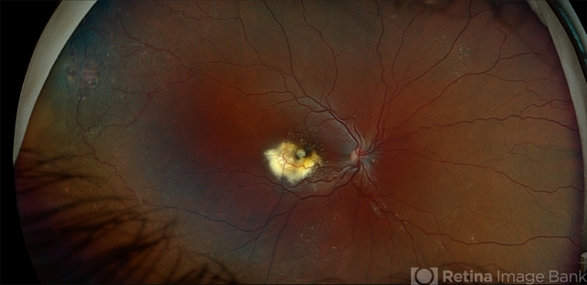
- Coat's disease
- Photographer
- Kimberly Wakester, COA
- Imaging device
-
Fundus camera
Optos California - Description
- Fundus photographs of an 7-year-old boy with Coat's Disease in the right eye. There is subfoveal lipid end scarring in the macula and "light bulb" type telangiectasias temporally noted on exam and shown in Optos color photos. FA findings show anastomoses, capillary dropout, and "light bulb" type telangiectasias temporally with mild late leakage. Patient will be monitored at this time and have repeat imaging in 4 months.


 Vasculitis")