Search results (216 results)
-
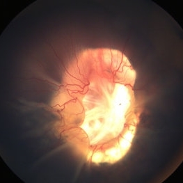
 Coats' Disease
Apr 27 2018 by Brenda Fallas
3-year-old boy with unilateral Coats' Disease fundus photo.
Photographer: Brenda Fallas, Bascom Palmer Eye Institute, Miami, FL
Imaging device: Retcam III 130 degree lens
Condition/keywords: Coats' disease, color fundus photograph, retinal telangiectasia
-
 Retinoblastoma
Apr 27 2018 by Brenda Fallas
2-year-old boy with stage D+ retinoblastoma of the right eye.
Photographer: Brenda Fallas, Bascom Palmer Eye Institute, Miami, FL
Imaging device: RETCAM III 130 degree lens montage
Condition/keywords: tumor, tumor seeding
-
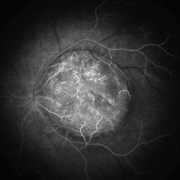
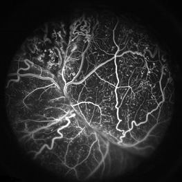 Coats' Disease FA
Apr 27 2018 by Brenda Fallas
3-year-old boy with unilateral Coats' Disease FA photo.
Photographer: Brenda Fallas, Bascom Palmer Eye Institute, Miami, FL
Imaging device: Retcam III 130 degree lens
Condition/keywords: Coats' disease, FA early phase, fluorescein angiogram (FA), retinal telangiectasia
-
 Coats' Disease
Apr 30 2020 by Giselle DeOliveira
Color Photograph of 20-month-old male infant.
Photographer: Giselle DeOliveira, University of Miami, Bascom Palmer Eye Institute
Imaging device: Retcam III
Condition/keywords: Coats' disease
-
 Retinal Detachment
Apr 30 2020 by Giselle DeOliveira
External / Gonio Photograph of 13-month old male infant with retinopathy of prematurity, retinal detachment and Cohen syndrome.
Photographer: Giselle DeOliveira, University of Miami, Bascom Palmer Eye Institute
Imaging device: Retcam III
-
 Cohen Syndrome Retinal Detachment
Apr 30 2020 by Giselle DeOliveira
Gonio Photograph of 13-month infant male with retinal detachment, retinopathy of prematurity and Cohen Syndrome
Photographer: Giselle DeOliveira, University of Miami, Bascom Palmer Eye Institute
Imaging device: Retcam III
Condition/keywords: retinopathy of prematurity (ROP)
-
 Retinoblastoma
Apr 23 2021 by Giselle DeOliveira
5 week old baby girl born with retinoblastoma.
Photographer: Giselle DeOliveira, Bascom Palmer Eye Institute, Miami ,Fl
Imaging device: Retcam
Condition/keywords: retinoblastoma
-
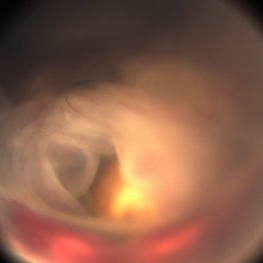
 Retinal Detachment Associated with Coloboma
Aug 23 2020 by Noy Ashkenazy, MD, MS
Fundus photograph of a 2-year-old boy with a history of CHARGE syndrome. The image nicely illustrates a retinal detachment associated with a congenital coloboma.
Photographer: Giselle DeOliveira
Imaging device: Retcam III
Condition/keywords: CHARGE syndrome, chronic retinal detachment, coloboma, pediatric retina
-
 Stage IVB
Oct 9 2012 by Audina M. Berrocal, MD FASRS
Progression of ROP to Stage IVB despite laser treatment.
Photographer: Ditte Hess CRA, BPEI
Imaging device: RetCam Digital Imaging
Condition/keywords: retinopathy of prematurity (ROP)
-
 Aggressive Posterior Retinopathy of Prematurity with Macular Hemorrhage
Oct 9 2012 by Audina M. Berrocal, MD FASRS
APROP with multiple pre-retinal hemorrhages
Photographer: Ditte Hess CRA, BPEI
Imaging device: RETCAM
Condition/keywords: macular hemorrhage, retinopathy of prematurity (ROP)
-
 Cat Eye Syndrome
Feb 11 2020 by Sophia El Hamichi, MD
A 3-year-old female with cat eye syndrome including iris, chorioretinal and optic nerve colobomas. Note the CNV temporally to the optic nerve coloboma (blue arrows)
Photographer: Giselle De Oliveira, Bascom Palmer Eye Institute, Miami
Imaging device: RetCam
Condition/keywords: cat eye syndrome, chorioretinal coloboma, choroidal neovascularization (CNV), coloboma, coloboma of optic disc, optic nerve coloboma
-
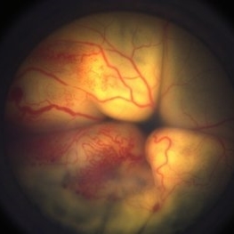
 Coats' Disease - Stage 3A
Aug 21 2019 by Victor M Villegas, MD
Coats' Disease - stage 3A.
Condition/keywords: abnormal retina, Coats' disease, diffuse lipid exudate, edema, foveal hard exudates, pediatic retina, retcam, retinal angioma
-
 Coats' Disease With Exudative Retinal Detachment and Retinal Macrocyst
Dec 9 2019 by Sophia El Hamichi, MD
A 3-year-old male with a presentation of a complex Coats' disease in the left eye with exudative retinal detachment, abnormal telangiectatic vasculature, and inferotemporal retinal macrocyst/retinoschisis.
Photographer: Abby Orcutt-Hayes, Murray Ocular Oncology and Retina
Imaging device: RetCam
Condition/keywords: Coats' disease, exudative detachment, montage, retinal macrocyst
-
 Retinopathy of Prematurity Stage 4a
Sep 7 2013 by Maria Ana Martinez-Castellanos, MD
Retinopathy of prematurity stage 4a.
Photographer: Maria A. Martinez-Castellanos. Asociacion para Evitar la Ceguera en Mexico
Imaging device: RetCam II
Condition/keywords: retinopathy of prematurity (ROP), retinopathy of prematurity stage 4a
-
 Aggressive Posterior Retinopathy of Prematurity with Macular Hemorrhage
Oct 9 2012 by Audina M. Berrocal, MD FASRS
Aggressive posterior Type 1 ROP
Photographer: Ditte Hess CRA, BPEI
Imaging device: RETCAM
Condition/keywords: aggressive posterior retinopathy of prematurity (APROP), macular hemorrhage, retinopathy of prematurity (ROP)
-
 Chorioretinal Coloboma with Retinal Detachment
Dec 5 2020 by Niloofar Piri, MD
14-year-old female with 1q21.1 microdeletion syndrome and behavioral, intellectual, and systemic abnormalities, including congenital microcornea, iris coloboma, and chorioretinal and optic nerve coloboma presented with decreased vision. Right eye fundus taken with RetCam shows coloboma with retinal detachment. (Left eye showed white cataract with funnel RD on B-scan).
Photographer: Niloofar Piri MD, Douglas Snyder MD
Condition/keywords: chorioretinal coloboma, optic nerve coloboma
-
 Eales Disease
Apr 3 2019 by Paola Brito, MD
8-year-old girl with positive Matoux test. She received laser in nasal retina. Peripheral vein occlusion, ischemic areas and neovascularization.
Photographer: Paola Brito, Hospital de la Luz, Mexico
Imaging device: retcam
Condition/keywords: Eales disease
-
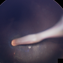
 Persistent Fetal Vasculature
Oct 18 2018 by Sengul Ozdek, MD, FEBO, FASRS
Fundus photograph of a 2-year-old girl who had already been operated elsewhere for congenital cataract before. Note the hyaloid vessels coming from ONH to the posterior capsule.
Photographer: Sengul Ozdek, Gazi University, School of Medicine.
Imaging device: RetCam
Condition/keywords: persistent fetal vasculature (PFV)
-
 Persistent Fetal Vasculature
May 8 2018 by Audina M. Berrocal, MD FASRS
Retrolental filling in PFV.
Photographer: Brenda Fallas
Imaging device: RETCAM FA
Condition/keywords: persistent fetal vasculature (PFV)
-
 Retinal Cyst
Aug 14 2020 by Noy Ashkenazy, MD, MS
Fundus photograph of a 13-year-old male with a chronic retinal detachment following a penetrating ocular trauma. There is a retinal cyst and proliferative vitreoretinopathy.
Photographer: Giselle DeOliveira
Imaging device: Retcam III
Condition/keywords: chronic retinal detachment, proliferative vitreoretinopathy (PVR), retinal cyst
-
 Retinal Fold Angiography
Feb 9 2017 by Dominic M Buzzacco, MD
Late phase angiogram of 10-month-old male with congenital retinal fold. Contralateral eye had normal angiography.
Photographer: Dominic M Buzzacco MD, Midwest Retina
Imaging device: Retcam 3
Condition/keywords: retinal fold
-
 Retinoblastoma
Sep 13 2013 by Maria Ana Martinez-Castellanos, MD
Fundus photograph, fluorescein angiography and OCT of the macula and of the tumor of a 2-years-old boy with retinoblastoma.
Photographer: Maria A. Martinez-Castellanos. Asociacion para Evitar la Ceguera en Mexico
Imaging device: RetCAm II
Condition/keywords: leakage, optical coherence tomography (OCT), pediatric tumor, retinoblastoma
-
 Retinoblastoma
Apr 23 2021 by Giselle DeOliveira
Angiography of retinoblastoma in 5 week old baby girl.
Photographer: Giselle DeOliveira, Bascom Palmer Eye Institute, Miami ,Fl
Imaging device: Retcam
Condition/keywords: retinoblastoma
-
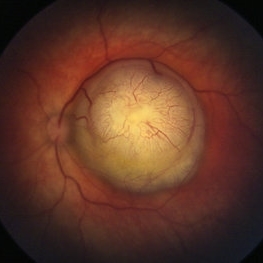
 Retinoblastoma Type 2 Regression After Chemo and Laser
Apr 17 2014 by Susanna S. Park, MD, PhD
Retcam fundus photograph of a 2-year-old boy with history of bilateral Group D retinoblastoma completing 6 cycles of systemic chemotherapy and retinal laser and cryotherapy with residual regressing posterior pole tumor showing predominantly type 2 regression. Pigmented rim shows scarring from prior diode and argon laser treatments.
Photographer: Ellen Redenbo, University of California Davis Eye Center
Condition/keywords: retina, retinoblastoma, type 2 regression
-
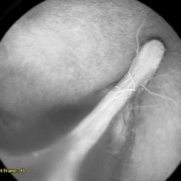
 Retinopathy of Prematurity
Feb 18 2015 by Andrea Arriola-Lopez, MD MSc
Closed funnel attached to posterior lens capsule.
Photographer: Andrea Elizabeth Arriola López, MSc. Asociación para Evitar la Ceguera, I.A.P. México D.F.
Imaging device: RetCam II
Condition/keywords: retinopathy of prematurity (ROP), stage 5
 Loading…