Search results (216 results)
-
 Retinopathy of Prematurity
Oct 27 2025 by Anjana Mirajkar, MS Ophthalmology
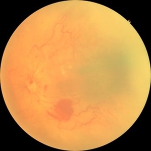
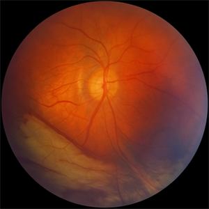
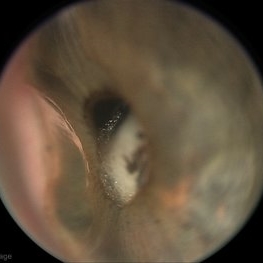
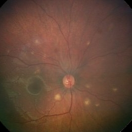
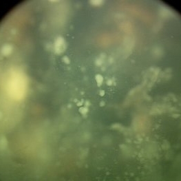
Fundus photograph of a premature baby showing flat neovascularization with looping of the vessels with bleed in zone 1/2 with plus disease suggestive of A-ROP.
Photographer: Dr. Anjana Mirajkar- HV desai eye hospital ,Pune
Imaging device: Retcam
Condition/keywords: aggressive posterior retinopathy of prematurity (APROP)
-
 Retinopathy of Prematurity
Oct 26 2025 by Anjana Mirajkar, MS Ophthalmology
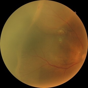
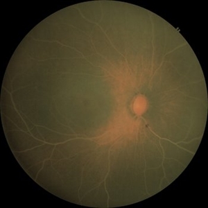
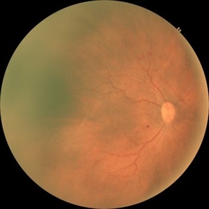
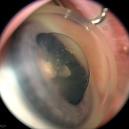
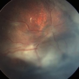
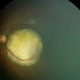
Fundus photograph of right eye of premature baby showing stage 3 in zone 2 posterior.
Photographer: Dr. Anjana Mirajkar- HV desai eye hospital ,Pune
Imaging device: Retcam
Condition/keywords: retinopathy of prematurity (ROP), stage 3
-
 Retinopathy of Prematurity
Oct 26 2025 by Anjana Mirajkar, MS Ophthalmology
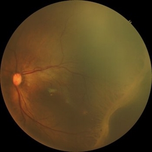
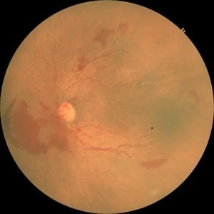
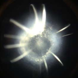
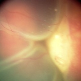
Fundus photograph of a left eye of a premature baby showing stage 3 in zone 2 posterior.
Photographer: Dr. Anjana Mirajkar- HV desai eye hospital ,Pune
Imaging device: Retcam
Condition/keywords: retinopathy of prematurity (ROP), retinopathy of prematurity stage 3
-
 Retinopathy of Prematurity
Oct 26 2025 by Anjana Mirajkar, MS Ophthalmology
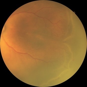
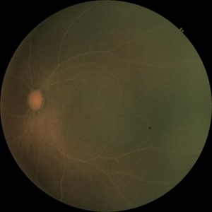
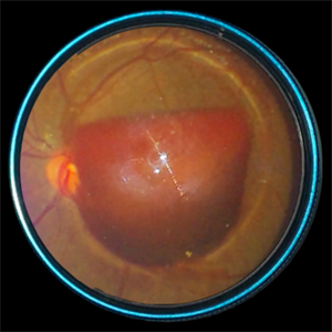
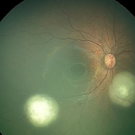
Fundus photograph of left eye premature baby having stage 3 in zone 2A with a secondary notch.
Photographer: Dr. Anjana Mirajkar- HV Desai eye hospital ,Pune
Imaging device: retcam
Condition/keywords: retinopathy of prematurity (ROP), stage 3
-
 Traction in Progression
Aug 6 2025 by Claudio Brancato, MD
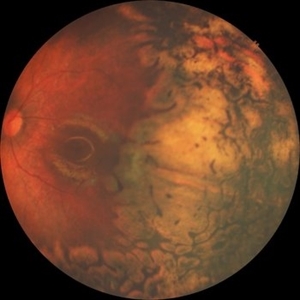
This image captures stage 4A Retinopathy of Prematurity, showing partial retinal detachment sparing the macula. The elevated retina and fibrous ridge indicate tractional forces secondary to extraretinal neovascularization. A striking representation of disease evolution, poised between reversibility and vision loss.
Photographer: Gregorio Lo Giudice, ARNAS Civico Hospital, Palermo, Italy
Imaging device: RETCAM 3 (enhanced via IA)
Condition/keywords: retinopathy of prematurity
-
 Lipemia Retinalis
Jun 3 2025 by Anjana Mirajkar, MS Ophthalmology
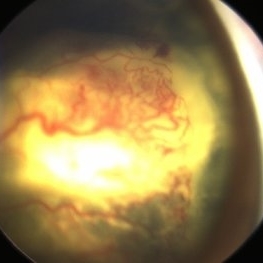
A fundus photograph of OD a premature baby of GA 34 weeks Birth weight - 1700gms,showing creamy colored blood vessels involving the entire posterior pole suggestive of severe changes of lipemia retinalis.
Photographer: Dhananjay Optometrist-H.V.Desai eye hospital, Pune
Imaging device: Retcam
Condition/keywords: lipemia retinalis
-
 Aggressive ROP
Jun 3 2025 by Anjana Mirajkar, MS Ophthalmology
Fundus photograph of OS of a premature baby of GA 29+2, Birth weight of 1325gms and Post menstrual age of 34+2, showing tortuosity and dilatation of vessels with looping in Zone 1 posterior with large pre retinal bleed nasal to disc suggestive of A-ROP.
Photographer: Vishnu Gaikwad- Optometrist H.V .Desai eye hospital, Pune
Imaging device: Retcam
Condition/keywords: aggressive posterior retinopathy of prematurity (APROP)
-
 Lipemia Retinalis
Jun 3 2025 by Anjana Mirajkar, MS Ophthalmology
A fundus photograph of OS a premature baby of GA 34 weeks Birth weight - 1700gms,showing creamy colored blood vessels involving the entire posterior pole suggestive of severe changes of lipemia retinalis.
Photographer: Dhananjay H.V.Desai eye hospital, Pune
Imaging device: Retcam
Condition/keywords: Lipemia
-
 Aggressive ROP
Jun 3 2025 by Anjana Mirajkar, MS Ophthalmology
Fundus photograph of OD of a premature baby of GA 29+2, Birth weight of 1325gms and Post menstrual age of 34+2, showing tortuosity and dilatation of vessels with looping in Zone 1 posterior suggestive of A-ROP.
Photographer: Vishnu Gaikwad- Optometrist H.V .Desai eye hospital, Pune
Imaging device: Retcam
Condition/keywords: aggresive retinopathy of prematurity
-
 Advance Coats' Disease
Feb 15 2025 by Theinchai Pasurakul, MD
From the fundus image, the peripheral retina exhibits telangiectatic vessels accompanied by light bulb aneurysms at their terminal ends.
Photographer: Michael J. Shapiro MD, Advocate Lutheran General Hospital, Des Plaines
Imaging device: Retcam
Condition/keywords: Coats' disease, light-bulb aneurysms
-
 Oval Pigmented Vitreous Cyst
Nov 27 2024 by Xinyu Zhao
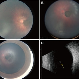
An 8-month-old infant was found to have a brown object in the left vitreous during a fundus screening. A wide-field digital retinal camera (RetCam) revealed a pigmented, non-transparent, freely floating, oval cystic lesion in the vitreous, measuring 2 disc diameters (Figures A-D). The cyst appeared cloudy when focused on the retina (Figure A) but was clearly defined in the vitreous (Figure B). Ultrasound showed a well-defined hyperreflective structure with a hyporeflective lumen (Figure D, indicated by the yellow arrow). A diagnosis of a vitreous pigment cyst, rare in infants, was made. Long-term follow-up is necessary to monitor changes affecting the infant’s vision.
Photographer: Xinyu Zhao, Shenzhen Eye Hospital, Shenzhen, China
Imaging device: RetCam
Condition/keywords: infant, vitreous cyst
-
 Fight for Sight
Mar 26 2024 by Tushar Agrawal
Fundus photograph showing 28 weeker APROP; regressed well after ROP Laser photocoagulation as seen at age 3 months.
Imaging device: Retcam neo
Condition/keywords: aggressive posterior retinopathy of prematurity (APROP), pediatric retina, retinopathy of prematurity (ROP)
-
 Retinal Detachment with Oil
Jun 14 2023 by Giselle DeOliveira
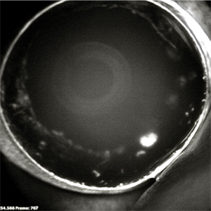
Fundus photograph of 16 year old girl with retinal detachment with oil from the angle view
Photographer: Giselle De Oliveira, University of Miami , Bascom Palmer Eye Institute
Imaging device: Envision Retcam 3
Condition/keywords: oil
-
 PFV
PFV
Jun 14 2023 by Giselle DeOliveira
Fundus photograph of a 6 week old girl with persistent fetal vasculature , view from the angle
Photographer: Giselle De Oliveira, University of Miami, Bascom Palmer Eye Institute
Imaging device: Envision Retcam 3
Condition/keywords: persistent fetal vasculature (PFV)
-
 Congenital cataract
Jun 14 2023 by Wenting Zhang
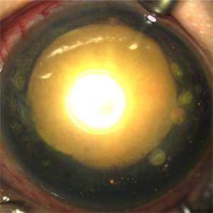
A two-year-old girl presents with a unilateral congenital cataract. The opacity appears to be mostly nuclear but with “riders” spreading into the adjacent cortex.
Photographer: Wenting Zhang, Department of Ophthalmology, Xin Hua Hospital Affiliated to Shanghai Jiao Tong University School of Medicine
Imaging device: Retcam III (Clarity Medical Systems, Pleasanton, CA, USA)
Condition/keywords: cataract
-
 SUBHYALOID HEMORRHAGE
Jul 27 2022 by Prithvi Chandrakanth
A 54-year-old male presented with sudden diminution of vision with visual acuity of 3/60 in the Left eye. visual acuity of the Right eye was 6/6, fundus examination revealed subhyaloid hemorrhage in the Left eye and normal fundus in the Right eye.
Photographer: Dr.Prithvi Chandrakanth, Department of Vitreoretinal Services, Aravind Eye Hospital, Coimbatore
Imaging device: TRASH TO TREASURE RETCAM - SMARTPHONE FUNDUS CAMERA
Condition/keywords: SHH, subhyaloid hemorrhage
-
 ROP-Zone-I-Stage-3-Plus
Jun 3 2022 by Dipak Nag, MBBS, FCPS, MSc, FRF
Fundus photograph of a child of gestational age 26 weeks and birth weight 1050 grams, shows dilatation and tortuosity of vessels in zone I, extra-retinal fibro-vascular proliferation, hemorrhage with huge peripheral avascular area.
Photographer: Dipak Nag, National Institute of Ophthalmology, Dhaka, Bangladesh
Imaging device: RetCam shuttle
Condition/keywords: retinopathy of prematurity (ROP), retinopathy of prematurity Plus disease, retinopathy of prematurity stage 3, retinopathy of prematurity zone I
-
 Miliary Tuberculosis
Mar 17 2022 by Franco Benvenuto, MD
Fundus photograph of a 9-month-old baby with hemophagocytic syndrome secondary to Tuberculosis infection.
Photographer: Franco Benvenuto, Universidad de Buenos Aires, Argentina
Imaging device: RetCam
Condition/keywords: ocular tuberculosis
-
 ROP 5A
Jan 24 2022 by Alexandre Grandinetti, MD, PhD
ROP retinal detachment.
Photographer: Alexandre Grandinetti
Imaging device: RetCam
Condition/keywords: retinopathy of prematurity (ROP), total retinal detachment
-
 Necrotic Multifocal Retinoblastoma Group E (ICRB) / cT3e (AJCC)
Jul 7 2021 by Linda A Cernichiaro- Espinosa, MD
A 3-year, 9-month-old male presented with unilateral advanced group E multifocal retinoblastoma cT3e (AJCC). Anterior seeding vascularized over the iris surface. Fluorescein angiogram fills the vascularized tumors. Aseptic orbital cellulitis, birefringent anterior segment crystals, cataract and dense vitritis are secondary to necrosis.
Photographer: Jose Oyervides-Alvarado MD
Imaging device: RetCam3
Condition/keywords: fluorescein angiogram (FA), pediatric tumor, retinoblastoma
-
 Necrotic Multifocal Retinoblastoma Group E (ICRB) / cT3e (AJCC)
Jul 7 2021 by Linda A Cernichiaro- Espinosa, MD
A 3-year, 9-month-old male presented with unilateral advanced group E multifocal retinoblastoma cT3e (AJCC). Anterior seeding vascularized over the iris surface. Fluorescein angiogram fills the vascularized tumors. Aseptic orbital cellulitis, birefringent anterior segment crystals, cataract and dense vitritis are secondary to necrosis.
Photographer: Jose Oyervides-Alvarado MD
Imaging device: RetCam3
Condition/keywords: retinoblastoma
-
 Vitreous Seeds
Jun 9 2021 by Thirumalesh Mochi Basavaraj, MD
Vitreous seeds in a case of retinoblastoma.
Photographer: Puttaswamy, Narayana Nethralaya, Bangalore
Imaging device: retcam
Condition/keywords: retinoblastoma
-
 Macular Retinoblastoma
Jun 9 2021 by Thirumalesh Mochi Basavaraj, MD
Macular retinoblastoma.
Photographer: Puttaswamy, Narayana Nethralaya
Imaging device: Retcam
Condition/keywords: tumor
-
 Retinoblastoma with Exudative Retinal Detachment
Jun 9 2021 by Thirumalesh Mochi Basavaraj, MD
A case of retinoblastoma with exudative retinal detachment and subretinal seeding.
Photographer: Puttaswamy, Narayan Nethralaya, Bangalore
Imaging device: Retcam
Condition/keywords: exudative retinal detachment
-
 Multifocal Retinoblastoma
Jun 9 2021 by Thirumalesh Mochi Basavaraj, MD
Inferotemporal tumor is blanched immediately post TTT, in comparison to the nasal tumor which has not received TTT.
Photographer: Puttaswamy, Narayana Nethralaya, Bangalore
Imaging device: Retcam
Condition/keywords: retinoblastoma, tumor
 Loading…