Search results (13 results)
-
 Retinoblastoma
Apr 23 2021 by Giselle DeOliveira
5 week old baby girl born with retinoblastoma.
Photographer: Giselle DeOliveira, Bascom Palmer Eye Institute, Miami ,Fl
Imaging device: Retcam
Condition/keywords: retinoblastoma
-
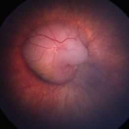
 Retinal Detachment Associated with Coloboma
Aug 23 2020 by Noy Ashkenazy, MD, MS
Fundus photograph of a 2-year-old boy with a history of CHARGE syndrome. The image nicely illustrates a retinal detachment associated with a congenital coloboma.
Photographer: Giselle DeOliveira
Imaging device: Retcam III
Condition/keywords: CHARGE syndrome, chronic retinal detachment, coloboma, pediatric retina
-
 Retinopathy of Prematurity S/P Laser Complications
Apr 30 2020 by Giselle DeOliveira
Gonio photograph of 13-year-old female with retinopathy of prematurity, s/p laser complications.
Photographer: Giselle DeOliveira
Imaging device: Retcam III
Condition/keywords: laser, retinopathy of prematurity (ROP)
-
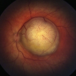
 Retinoblastoma
Apr 30 2020 by Giselle DeOliveira
Fundus photograph of an 17-month-old female infant with retinoblastoma over optic nerve.
Photographer: Giselle DeOliveira, University of Miami, Bascom Palmer Eye Institute
Imaging device: Retcam III
Condition/keywords: retinoblastoma
-
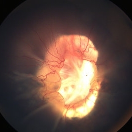
 Coats' Disease With Exudative Retinal Detachment and Retinal Macrocyst
Dec 9 2019 by Sophia El Hamichi, MD
A 3-year-old male with a presentation of a complex Coats' disease in the left eye with exudative retinal detachment, abnormal telangiectatic vasculature, and inferotemporal retinal macrocyst/retinoschisis.
Photographer: Abby Orcutt-Hayes, Murray Ocular Oncology and Retina
Imaging device: RetCam
Condition/keywords: Coats' disease, exudative detachment, montage, retinal macrocyst
-
 Coats' Disease - Stage 3A
Aug 21 2019 by Victor M Villegas, MD
Coats' Disease - stage 3A.
Condition/keywords: abnormal retina, Coats' disease, diffuse lipid exudate, edema, foveal hard exudates, pediatic retina, retcam, retinal angioma
-
 Persistent Fetal Vasculature
Oct 18 2018 by Sengul Ozdek, MD, FEBO, FASRS
Fundus photograph of a 2-year-old girl who had already been operated elsewhere for congenital cataract before. Note the hyaloid vessels coming from ONH to the posterior capsule.
Photographer: Sengul Ozdek, Gazi University, School of Medicine.
Imaging device: RetCam
Condition/keywords: persistent fetal vasculature (PFV)
-
 Retinal Cavernous Hemangioma
Nov 30 2018 by Brenda Fallas
2-year-old female with retinal cavernous hemangioma.
Photographer: Brenda Fallas
Imaging device: Retcam 130 lens
Condition/keywords: cluster of grapes, retinal angioma
-
 Coats' Disease
Apr 27 2018 by Brenda Fallas
3-year-old boy with unilateral Coats' Disease fundus photo.
Photographer: Brenda Fallas, Bascom Palmer Eye Institute, Miami, FL
Imaging device: Retcam III 130 degree lens
Condition/keywords: Coats' disease, color fundus photograph, retinal telangiectasia
-
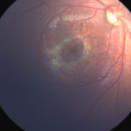 Congenital Zika Syndrome
Jun 29 2017 by Camila V Ventura, MD, PhD
Fundus image of an infant with congenital zika syndrome presenting with focal pigmentary changes in the macular region
Photographer: Camila Ventura, MD - Altino Ventura Foundation, Brazil
Imaging device: Retcam®
Condition/keywords: focal pigmentary changes
-
 FEVR with Knife-Like Fold
Jan 11 2014 by Caesar K. Luo, MD, FASRS
Retcam photograph of child with FEVR.
Photographer: Caesar Luo, Progressive Vision Institute, PA
Imaging device: RetCam
Condition/keywords: familial exudative vitreoretinopathy (FEVR)
-
 Aggressive Posterior Retinopathy of Prematurity (AP-ROP)
Nov 21 2013 by Rini Hersetyati, MD
This child with AP-ROP was born 31 weeks gestational age and 1700g birth weight. He was imaged at 38 weeks.
Photographer: Dr. Rini Hersetyati, Klinik Mata Nusantara, Jakarta, Indonesia
Imaging device: RetCam Fluorescein Angiography
Condition/keywords: aggressive posterior retinopathy of prematurity (APROP)
-
 Retinopathy of Prematurity Stage 4a
Sep 7 2013 by Maria Ana Martinez-Castellanos, MD
Retinopathy of prematurity stage 4a.
Photographer: Maria A. Martinez-Castellanos. Asociacion para Evitar la Ceguera en Mexico
Imaging device: RetCam II
Condition/keywords: retinopathy of prematurity (ROP), retinopathy of prematurity stage 4a
 Loading…