File number: 26964
Comments
-
 James B. Soque, CRA, OCT-C, COA, FOPS (May 17 2017)
The details of this late venous filling angiogram are alarming, the simply fantastic.
Thank you for your excellent submission Dr Bizzacco.
Sign in to comment.
 Initializing download.
-
 By Dominic M Buzzacco, MD
The Ohio State University Medical Center
Co-author(s): MaryLou McGregor MD, Nationwide Childrens Hospital, Columbus OH - Uploaded on Feb 9, 2017.
- Last modified by Caroline Bozell on Feb 10, 2017.
- Rating
- Appears in
- Miscellaneous
- Condition/keywords
- retinal fold
- Photographer
- Dominic M Buzzacco MD, Midwest Retina
- Imaging device
- Retcam 3
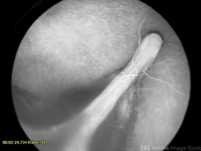
- Description
- Late phase angiogram of 10-month-old male with congenital retinal fold. Contralateral eye had normal angiography.


