Search results (216 results)
-
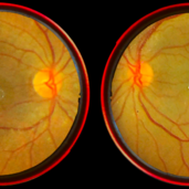
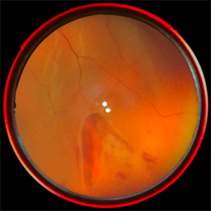
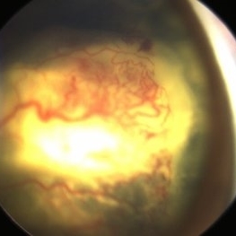 Advance Coats' Disease
Feb 15 2025 by Theinchai Pasurakul, MD
From the fundus image, the peripheral retina exhibits telangiectatic vessels accompanied by light bulb aneurysms at their terminal ends.
Photographer: Michael J. Shapiro MD, Advocate Lutheran General Hospital, Des Plaines
Imaging device: Retcam
Condition/keywords: Coats' disease, light-bulb aneurysms
-
 Aggressive Posterior Retinopathy of Prematurity
Oct 9 2012 by Audina M. Berrocal, MD FASRS
Aggressive posterior Type 1 ROP with bleeding from regression of the posterior hyaloid artery
Photographer: Ditte Hess CRA, BPEI
Imaging device: RETCAM
Condition/keywords: retinopathy of prematurity (ROP)
-
 Aggressive Posterior Retinopathy of Prematurity with Macular Hemorrhage
Oct 9 2012 by Audina M. Berrocal, MD FASRS
Aggressive posterior Type 1 ROP
Photographer: Ditte Hess CRA, BPEI
Imaging device: RETCAM
Condition/keywords: aggressive posterior retinopathy of prematurity (APROP), macular hemorrhage, retinopathy of prematurity (ROP)
-
 Aggressive Posterior Retinopathy of Prematurity with Macular Hemorrhage
Oct 9 2012 by Audina M. Berrocal, MD FASRS
APROP with multiple pre-retinal hemorrhages
Photographer: Ditte Hess CRA, BPEI
Imaging device: RETCAM
Condition/keywords: macular hemorrhage, retinopathy of prematurity (ROP)
-
 Aggressive Posterior ROP With Laser
Aug 18 2018 by Anna L. Ells, MD, FRCS(C)
Plus disease and aggressive posterior ROP, immediately following laser to peripheral avascular retina.
Photographer: Anna Ells
Imaging device: RetCam
Condition/keywords: aggressive posterior retinopathy of prematurity (APROP)
-
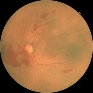
 Aggressive ROP
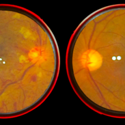
Jun 3 2025 by Anjana Mirajkar, MS Ophthalmology
Fundus photograph of OD of a premature baby of GA 29+2, Birth weight of 1325gms and Post menstrual age of 34+2, showing tortuosity and dilatation of vessels with looping in Zone 1 posterior suggestive of A-ROP.
Photographer: Vishnu Gaikwad- Optometrist H.V .Desai eye hospital, Pune
Imaging device: Retcam
Condition/keywords: aggresive retinopathy of prematurity
-
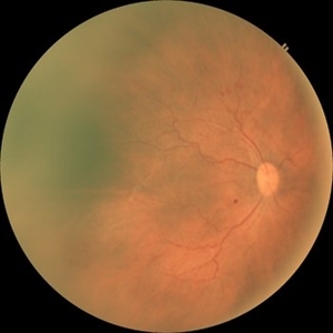
 Aggressive ROP
Jun 3 2025 by Anjana Mirajkar, MS Ophthalmology
Fundus photograph of OS of a premature baby of GA 29+2, Birth weight of 1325gms and Post menstrual age of 34+2, showing tortuosity and dilatation of vessels with looping in Zone 1 posterior with large pre retinal bleed nasal to disc suggestive of A-ROP.
Photographer: Vishnu Gaikwad- Optometrist H.V .Desai eye hospital, Pune
Imaging device: Retcam
Condition/keywords: aggressive posterior retinopathy of prematurity (APROP)
-
 Bear Tracks - Smartphone Fundus Image
Dec 27 2018 by Prithvi Chandrakanth
Male 43-years-old, BCVA BE 6/6, asymptomatic, Fundus RE multiple hyperpigmented circular/oval well defined lesions present inferonasally to the optic disc.
Photographer: Dr.Prithvi Chandrakanth, Dr.Chandrakanth Malabar Nethralaya, Kozhikode
Imaging device: Trash To Treasure Retcam - Smartphone Fundus Camers
Condition/keywords: bear tracks, congenital hypertrophy of the retinal pigment epithelium (CHRPE), retcam, smartphone fundus photography
-
 Branch Retinal Vein Occlusion
Feb 5 2020 by Prithvi Chandrakanth
54-year-old female presented with blurring of vision in the left eye , anterior segment evaluation revealed immature cataract in both the eye and fundus evaluation revealed normal picture in the right eye and branch retinal vein occlusion in the left eye.
Photographer: Dr.PRITHVI CHANDRAKANTH, ARAVIND EYE HOSPITAL, UDUMALPET
Imaging device: TRASH TO TREASURE RETCAM
Condition/keywords: branch retinal vein occlusion (BRVO), retcam, smartphone fundus photography
-
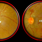
 Cat Eye Syndrome
Feb 11 2020 by Sophia El Hamichi, MD
A 3-year-old female with cat eye syndrome including iris, chorioretinal and optic nerve colobomas. Note the CNV temporally to the optic nerve coloboma (blue arrows)
Photographer: Giselle De Oliveira, Bascom Palmer Eye Institute, Miami
Imaging device: RetCam
Condition/keywords: cat eye syndrome, chorioretinal coloboma, choroidal neovascularization (CNV), coloboma, coloboma of optic disc, optic nerve coloboma
-
 Central Retinal Artery Occlusion
Jan 13 2020 by Prithvi Chandrakanth
37-years-old male with complaints of sudden diminution of vision in the left eye for the past three days. Fundus examination revealed pale retina in the left eye with cherry red spot and normal fundus picture in right eye.
Photographer: DR.PRITHVI CHANDRAKANTH, ARAVIND EYE HOSPITAL, UDUMALPET
Imaging device: TRASH TO TREASURE RETCAM
Condition/keywords: central retinal artery occlusion (CRAO), cherry red spot, retcam, smartphone fundus photography
-
 Central Retinal Vein Occlusion
Jan 26 2020 by Prithvi Chandrakanth
Transient visual obscuration in a 52-year-old male patient in his right eye, fundus examination revealed dot, blot and flame shaped hemorrhage with cotton wool patches.
Photographer: Dr.PRITHVI CHANDRAKANTH, ARAVIND EYE HOSPITAL, UDUMALPET
Imaging device: TRASH TO TREASURE RETCAM
Condition/keywords: central retinal vein occlusion (CRVO), retcam, smartphone fundus photography
-
 Coats With Exudative Retinal Detachment
Jan 11 2014 by Caesar K. Luo, MD, FASRS
Retcam photograph of child with Coat's disease with extensive exudation and large exudative retinal detachment.
Photographer: Caesar Luo, Progressive Vision Institute, PA
Imaging device: RetCam
-
 Coats' Disease - Stage 3A
Aug 21 2019 by Victor M Villegas, MD
Coats' Disease - stage 3A.
Condition/keywords: abnormal retina, Coats' disease, diffuse lipid exudate, edema, foveal hard exudates, pediatic retina, retcam, retinal angioma
-
 Coats' Disease With Exudative Retinal Detachment and Retinal Macrocyst
Dec 9 2019 by Sophia El Hamichi, MD
A 3-year-old male with a presentation of a complex Coats' disease in the left eye with exudative retinal detachment, abnormal telangiectatic vasculature, and inferotemporal retinal macrocyst/retinoschisis.
Photographer: Abby Orcutt-Hayes, Murray Ocular Oncology and Retina
Imaging device: RetCam
Condition/keywords: Coats' disease, exudative detachment, montage, retinal macrocyst
-
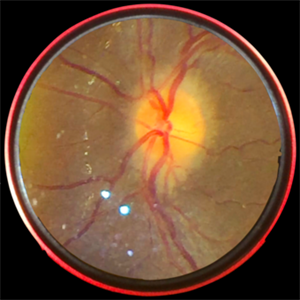 Disc Edema
Oct 16 2019 by Prithvi Chandrakanth
26-year-old male, presented with defective vision, on examination his color vision, red desaturation test were reduced. Fundus examination revealed edematous halo around the disc suggesting progressive optic disc edema.
Photographer: Dr.Prithvi Chandrakanth, Dr.Chandrakanth Malabar Nethralaya, Kozhikode, India
Imaging device: TRASH TO TREASURE RETCAM
Condition/keywords: frisen grade 2, optic disc edema, retcam, smartphone fundus photography
-
 Eales Disease
Apr 3 2019 by Paola Brito, MD
8-year-old girl with positive Matoux test. She received laser in nasal retina. Peripheral vein occlusion, ischemic areas and neovascularization.
Photographer: Paola Brito, Hospital de la Luz, Mexico
Imaging device: retcam
Condition/keywords: Eales disease
-
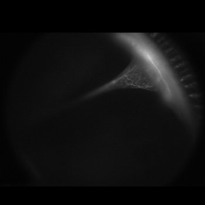 FEVR
FEVR
Apr 29 2021 by Giselle DeOliveira
Fluorescein image of 2-year-old female with familial exudative vitrepretinpoathy detachment touching the lens.
Photographer: Giselle DeOliveira
Imaging device: Retcam
Condition/keywords: familial exudative vitreoretinopathy (FEVR)
-
 FEVR Fluorescein
Jan 11 2014 by Caesar K. Luo, MD, FASRS
Retcam fluorescein angiography of child with FEVR. Peripheral nonperfusion and knife-like fold
Photographer: Caesar Luo, Progressive Vision Institute, PA
Imaging device: RetCam
Condition/keywords: familial exudative vitreoretinopathy (FEVR)
-
 FEVR with Knife-Like Fold
Jan 11 2014 by Caesar K. Luo, MD, FASRS
Retcam photograph of child with FEVR.
Photographer: Caesar Luo, Progressive Vision Institute, PA
Imaging device: RetCam
Condition/keywords: familial exudative vitreoretinopathy (FEVR)
-
 High Magnification of Stage 3 Neovascularization
Aug 18 2018 by Anna L. Ells, MD, FRCS(C)
High magnification highlighting neovascularization of stage 3 ROP. Note the "popcorn" just posterior to the neovascularization.
Photographer: Leslie Mackeen
Imaging device: Retcam
Condition/keywords: retinopathy of prematurity stage 3
-
 High Magnification of Stage 3 Neovascularization in ROP
Aug 18 2018 by Anna L. Ells, MD, FRCS(C)
High magnification of stage 3 neovascularization in ROP.
Photographer: Leslie MacKeen
Imaging device: RetCam
Condition/keywords: retinopathy of prematurity stage 3
-
 Horse Shoe Retinal Tear
Feb 5 2020 by Prithvi Chandrakanth
46-year-old male presented with blurring of vision in the right eye, anterior segment in the right eye was normal with positive staffers sign and fundus picture consisting of horse shoe tear in the peripheral retina.
Photographer: Dr. PRITHVI CHANDRAKANTH, ARAVIND EYE HOSPITAL, UDUMALPET
Imaging device: TRASH TO TREASURE RETCAM
Condition/keywords: retcam, smartphone fundus photography
-
 Juvenile X-linked Retinoschisis
Jan 11 2014 by Caesar K. Luo, MD, FASRS
Retcam photograph of child with JXLRS.
Photographer: Caesar Luo, Progressive Vision Institute, PA
Imaging device: RetCam
Condition/keywords: juvenile retinoschisis
-
 Lamellar Congenital Cataracts
Aug 24 2020 by Sophia El Hamichi, MD
A 4-month-old female with family history of congenital cataract was referred for bilateral congenital cataract with nystagmus and torticoli. During the ophthalmological exam we were able to access the fundus and perform a fluorescein angiogram through the clear ring surrounding the lens opacity. Retina evaluation showed no abnormalities.
Photographer: Abby Orcutt-Hayes, Murray Ocular Oncology and Retina
Imaging device: RetCam
Condition/keywords: congenital cataract, lamellar cataract, nystagmus
 Loading…