Search results (216 results)
-
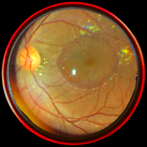
 Stage 5 Retinopathy of Prematurity (ROP)
Oct 9 2012 by Audina M. Berrocal, MD FASRS
Advanced APROP with Stage 5 and vascularly active.
Photographer: Ditte Hess CRA, BPEI
Imaging device: RETCAM
Condition/keywords: retinopathy of prematurity (ROP), stage 5
-
 Aggressive Posterior Retinopathy of Prematurity with Macular Hemorrhage
Oct 9 2012 by Audina M. Berrocal, MD FASRS
APROP with multiple pre-retinal hemorrhages
Photographer: Ditte Hess CRA, BPEI
Imaging device: RETCAM
Condition/keywords: macular hemorrhage, retinopathy of prematurity (ROP)
-
 Retinoblastoma with Multiple Vitreous Seeding
Oct 9 2012 by Audina M. Berrocal, MD FASRS
Retinoblastoma with vitreous seeding.
Photographer: Ditte Hess CRA, BPEI
Imaging device: RETCAM
Condition/keywords: retinoblastoma, vitreous seeding
-
 X-linked Retinoschisis (XLRS)
Oct 9 2012 by Audina M. Berrocal, MD FASRS
6-week-old baby with XLRS
Photographer: Ditte Hess CRA, BPEI
Imaging device: RETCAM/OIS
Condition/keywords: retinoschisis, x-linked retinoschisis (XLRS)
-
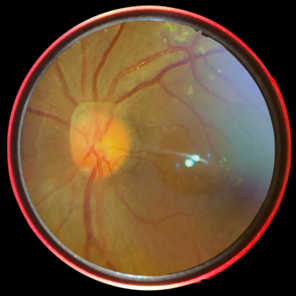
 Aggressive Posterior Retinopathy of Prematurity
Oct 9 2012 by Audina M. Berrocal, MD FASRS
Aggressive posterior Type 1 ROP with bleeding from regression of the posterior hyaloid artery
Photographer: Ditte Hess CRA, BPEI
Imaging device: RETCAM
Condition/keywords: retinopathy of prematurity (ROP)
-
 Skip Area in Retinopathy of Pretmaturity
Oct 9 2012 by Audina M. Berrocal, MD FASRS
Skip area in ROP after laser treatment.
Photographer: Ditte Hess CRA, BPEI
Imaging device: RETCAM
Condition/keywords: laser, retinopathy of prematurity (ROP)
-
 Stage IVB
Oct 9 2012 by Audina M. Berrocal, MD FASRS
Progression of ROP to Stage IVB despite laser treatment.
Photographer: Ditte Hess CRA, BPEI
Imaging device: RetCam Digital Imaging
Condition/keywords: retinopathy of prematurity (ROP)
-
 Persistent Fetal Vasculature (PFV)
Oct 9 2012 by Audina M. Berrocal, MD FASRS
Baby with PFV
Photographer: Ditte Hess CRA, BPEI
Imaging device: RETCAM
Condition/keywords: persistent fetal vasculature (PFV)
-
 Aggressive Posterior Retinopathy of Prematurity (AP-ROP)
Feb 8 2013 by Sjakon G Tahija, MD
Fundus photograph of a premature neonate with APROP.
Photographer: Rini Hersetyati, M.D.; Klinik Mata Nusantara, Jakarta, Indonesia
Imaging device: RetCam II - Clarity Medical Systems, U.S.A.
Condition/keywords: aggressive posterior retinopathy of prematurity (APROP)
-
 Stage 3 ROP
Aug 18 2018 by Anna L. Ells, MD, FRCS(C)
Left retinal image of premature infant born at 24 weeks gestational age, birthweight of 653 grams. Image taken at 38 weeks of post-menstrual age. Zone II, stage 3 ROP. Moderate plus disease. Good example of Type I ROP requiring treatment.
Photographer: Leslie Mackeen, Hospital for Sick Children, Toronto, Canada
Imaging device: RetCam
Condition/keywords: retinopathy of prematurity stage 3
-
 Aggressive Posterior Retinopathy of Prematurity with Macular Hemorrhage
Oct 9 2012 by Audina M. Berrocal, MD FASRS
Aggressive posterior Type 1 ROP
Photographer: Ditte Hess CRA, BPEI
Imaging device: RETCAM
Condition/keywords: aggressive posterior retinopathy of prematurity (APROP), macular hemorrhage, retinopathy of prematurity (ROP)
-
 Retinoblastoma Type 2 Regression After Chemo and Laser
Apr 17 2014 by Susanna S. Park, MD, PhD
Retcam fundus photograph of a 2-year-old boy with history of bilateral Group D retinoblastoma completing 6 cycles of systemic chemotherapy and retinal laser and cryotherapy with residual regressing posterior pole tumor showing predominantly type 2 regression. Pigmented rim shows scarring from prior diode and argon laser treatments.
Photographer: Ellen Redenbo, University of California Davis Eye Center
Condition/keywords: retina, retinoblastoma, type 2 regression
-
 Coats' Disease
Apr 27 2018 by Brenda Fallas
3-year-old boy with unilateral Coats' Disease fundus photo.
Photographer: Brenda Fallas, Bascom Palmer Eye Institute, Miami, FL
Imaging device: Retcam III 130 degree lens
Condition/keywords: Coats' disease, color fundus photograph, retinal telangiectasia
-
 Retinoblastoma
Apr 27 2018 by Brenda Fallas
2-year-old boy with stage D+ retinoblastoma of the right eye.
Photographer: Brenda Fallas, Bascom Palmer Eye Institute, Miami, FL
Imaging device: RETCAM III 130 degree lens montage
Condition/keywords: tumor, tumor seeding
-
 Shaken Baby Syndrome
Feb 27 2017 by Joana Rita de Medeiros Pinto
Fundus photograph of a 3-month-old baby with shaken baby syndrome.
Photographer: Joana Pinto, Hospital de Santa Maria, Lisbon
Imaging device: Retcam3
Condition/keywords: shaken baby syndrome
-
 Retinoblastoma
Sep 13 2013 by Maria Ana Martinez-Castellanos, MD
Fundus photograph, fluorescein angiography and OCT of the macula and of the tumor of a 2-years-old boy with retinoblastoma.
Photographer: Maria A. Martinez-Castellanos. Asociacion para Evitar la Ceguera en Mexico
Imaging device: RetCAm II
Condition/keywords: leakage, optical coherence tomography (OCT), pediatric tumor, retinoblastoma
-
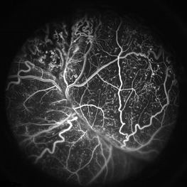
 Juvenile X-linked Retinoschisis
Jan 11 2014 by Caesar K. Luo, MD, FASRS
RetCam fluorescein angiography of child with JXLRS.
Photographer: Caesar Luo, Progressive Vision Institute, PA
Condition/keywords: juvenile retinoschisis
-
 Stage 4A Retinopathy of Prematurity
Nov 7 2013 by Maria Ana Martinez-Castellanos, MD
Nasal area of the left eye of a baby with ROP stage 4A.
Photographer: Maria A. Martinez-Castellanos. Asociacion para Evitar la Ceguera en Mexico
Imaging device: RetCam II
Condition/keywords: retinopathy of prematurity stage 4a, tractional retinal detachment
-
 Central Serous Chorioretinopathy : Smartphone Fundus Image
Dec 14 2018 by Prithvi Chandrakanth
A 42-year-old male with diminution of vision in the left eye since one week, uncorrected visual acuity was 6/18 improving with hyperopic lens to 6/9. H/o Metamorphopsia was present.
Photographer: Dr.Prithvi Chandrakanth, Dr.Chandrakanth Malabar Nethralaya, Kozhikode.
Imaging device: Trash To Treasure (T3) Retcam - Smartphone Fundus Camera
Condition/keywords: central serous chorioretinopathy (CSCR), smartphone fundus photography
-
 Persistent Fetal Vasculature (PFV)
Oct 9 2012 by Audina M. Berrocal, MD FASRS
Photographer: Ditte Hess CRA, BPEI
Imaging device: RETCAM
Condition/keywords: persistent fetal vasculature (PFV)
-
 Persistent Fetal Vasculature
Oct 18 2018 by Sengul Ozdek, MD, FEBO, FASRS
Fundus photograph of a 2-year-old girl who had already been operated elsewhere for congenital cataract before. Note the hyaloid vessels coming from ONH to the posterior capsule.
Photographer: Sengul Ozdek, Gazi University, School of Medicine.
Imaging device: RetCam
Condition/keywords: persistent fetal vasculature (PFV)
-
 Bergmeisters-Papilla : Smartphone Fundus Image
Dec 14 2018 by Prithvi Chandrakanth
Smartphone fundus photograph of a 33-year-old male with Bergmeister's papilla.
Photographer: Dr.Prithvi Chandrakanth, Dr.Chandrakanth Malabar Nethralaya, Kozhikode.
Imaging device: Trash To Treasure (T3) Retcam Smartphone Fundus Camera
Condition/keywords: Bergmeister's Papillae, smartphone fundus photography
-
 Coats' Disease FA
Apr 27 2018 by Brenda Fallas
3-year-old boy with unilateral Coats' Disease FA photo.
Photographer: Brenda Fallas, Bascom Palmer Eye Institute, Miami, FL
Imaging device: Retcam III 130 degree lens
Condition/keywords: Coats' disease, FA early phase, fluorescein angiogram (FA), retinal telangiectasia
-
 Retinoblastoma
Nov 7 2013 by Maria Ana Martinez-Castellanos, MD
Retinoblastoma in a 2-year-old boy.
Photographer: Maria A. Martinez-Castellanos. Asociacion para Evitar la Ceguera en Mexico
Imaging device: RetCamII
Condition/keywords: retinoblastoma
-
 Group D Retinoblastoma After Chemo and Laser
Apr 17 2014 by Susanna S. Park, MD, PhD
Retcam fundus photograph of a 2 year old boy with history of bilateral Group D retinoblastoma completing 6 cycles of systemic chemotherapy and retinal laser and cryotherapy with residual regressing posterior pole tumor showing type 3 (type 1 and 2) regression pattern. Some pigmented scarring and hemorrhage are also noted nasal to the disc from prior laser treatment of tumor.
Photographer: Ellen Redenbo
Condition/keywords: retina
 Loading…