Search results (333 results)
-
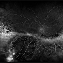
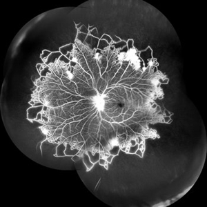 Vascular Non Perfusion in Takayasu Arteritis
Feb 6 2024 by SHILPI H NARNAWARE, ICO ( Retina) , FAICO ( Vitreo-Retina)
A case of 16 year-old female with combined RD in RE. Fundus examination & FFA revealed 360 degrees non-perfusion in periphery in non-symptomatic eye.
Photographer: Shilpi Narnaware, Sarakshi Netralaya , Nagpur, Maharashtra , India
Imaging device: Mirante ( by Nidek)
Condition/keywords: CNP areas, takayasu arteritis
-
 Benign Familial Fleck Retina
Nov 7 2018 by Vedang Shah
Flecks over the entire retinal mid-periphery and periphery of a 12-year-old male with no visual complaints.
Photographer: Dr. Vedang Shah
Imaging device: OPTOS IMAGING SYSTEM
Condition/keywords: fleck retinopathy
-
 Acute Retinal Necrosis (ARN)
Jul 3 2025 by Heitor Nogueira
Fundus photograph of an 63-year-old woman who reported unilateral visual acuity loss for 10 days associated with ocular pain. He presented conjunctival hyperemia with temporal and nasal nodular scleritis, anterior chamber reaction 2+/4+, Koeppe nodules, granulomatous PKs, vitreitis 2+/4+, multiple areas of vasculitis in the arcades and periphery, associated with hemorrhages and necrotizing retinitis in the temporal, inferior and nasal periphery. Positive serology for Herpes Virus
Photographer: Heitor Nogueira, Penido Burnier Institute, Campinas, São Paulo, Brazil
Imaging device: Optos Daytona
Condition/keywords: ARN complications, Herpes, progressive outer retinal necrosis (PORN), Uveitis
-
 CRVO
CRVO
Apr 22 2017 by Gabriel Costa Andrade, PhD
Panoramic retinography (Optos® California) of the right eye of a 48-year-old female patient with a history of low-vision in the right eye 2 months ago. At the exam presented visual acuity of 20/200 in the right eye and 20/20 in the left eye. Angiography shows diffuse perivascular leakage associated with areas of hypoperfusion in macula and periphery.
Photographer: Gabriel Andrade
Imaging device: Optos® California
Condition/keywords: central retinal vein occlusion (CRVO)
-
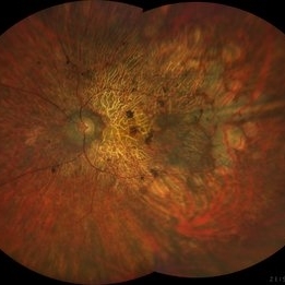 Rod Cone dystrophy
Nov 29 2022 by Niloofar Piri, MD
Fundus photograph of the left eye in a 58 yo male with rod cone dystrophy. He presented with night blindness and peripheral vision loss since youth and recent decrease in central vision for the past 10 years. Notice waxy pallor of the nerve, severe arterial narrowing and chorioretinal atrophy mainly around the arcades as well as posterior pole along with RPE hyperplastic changes and atrophy. RPE atrophy in midperiphery has coin shaped appearance. FAF has characteristic appearance (uploaded separately) He has one pathogenic variants of both CEP290 and PRPH2 genes.
Photographer: Sean Kelso, Saint Louis University
Condition/keywords: hereditary retinal deg, hereditary retinal dystrophy, Rod cone dystrophy
-
 Benign Idiopathic Hemorrhagic Retiopathy - RF Image
Feb 6 2013 by Roy Schwartz, MD
A healthy 31-year-old female presented with visual deterioration in her left eye. On examination thick macular retinal hemorrhages were seen, as well as some small minopr hemorrhages in the periphery. A workup for hypercoagulability was negative, and sources of emboli were not found. A diagnosis of a unilateral idiopathic hemorrhagic retinopathy was made.
Photographer: Galit Yair-Pur
Condition/keywords: benign idiopathic hemorrhagic retinopathy, red-free
-
 Coats' Disease
Feb 25 2021 by Niloofar Piri, MD
Collage color photo and FA image of the same patient with Coats' Disease demonstrating telangiectatic aneurysmal lesions in the temporal periphery, associated with hard exudate deposition posteriorly. FA (AV phase) demonstrating hyperfluorescent aneurysmal lesions as well as peripheral capillary non perfusion. Note the posterior hypofluorescence where the hard exudates are located.
Condition/keywords: Coats' disease, congenital retinal telangiectasis, retinal telangiectasia
-
 Coats' Disease
Feb 2 2021 by Niloofar Piri, MD
#4 Recirculation phase fluorescein angiography of the same patient demonstrating increased hyperfluorescence and leakage from abnormal vascular lesions in temporal periphery. Note the capillary non perfusion area anteriorly.
Condition/keywords: Coats' disease, Leber's miliary aneurysm
-
 Peripheral CNVM with Extensive Scarring
Oct 12 2019 by John S. King, MD
82-year-old white male with an acute loss of vision in the right eye was sent in to rule out a retinal detachment. Vision was 20/350; a dense VH was present, b-scan showed irregular areas of high reflectivity in the periphery that was c/w SRH. Peripherally, a few weeks later, there were areas that could be seen and were c/w peripheral CNVM (old and new). Anti-VEGF was administered. A month later vision was unchanged and patient wanted surgery to remove the VH. Pictured is one week since surgery; large peripheral scars are seen; diffuse areas of SR pigmentation is present; vitreous skirt present; and a few IRHs secondary to DR can be seen. He is currently 20/70 sc.
Photographer: Shelly Blair
Imaging device: Optos CA
Condition/keywords: choroidal neovascular membrane (CNVM), peripheral fundus lesion, vitreous blood
-
 Proliferative Sickle Cell Retinopathy
Jan 29 2021 by Olivia Rainey
Ultra-widefield fluorescein angiogram of a 24-year-old female with proliferative sickle cell retinopathy affecting her right eye. The physician's interpretation of the fluorescein shows seafan neovascularization superotemporally, AV anastomeses, and good peripheral laser. He performed scatter PRP OD on 12/2/2020 to nonperfusion in temporal far periphery. The patient's 12/2020 Hb electrophoresis came back showing Hb SC (rather than sickle cell trait). Patient was born at full term, but she reports that her mother used drugs while pregnant with the patient. The patient also mentioned that her niece has full sickle cell disease and her grandmother, mother, and sibling all have condition on the sickle cell spectrum.
Photographer: Olivia Rainey, OCT-C, COA
Imaging device: Optos California
Condition/keywords: fluorescein angiogram (FA), fluorescein leakage, neovascularization (NV), neovascularization elsewhere (NVE), Optos, sea fan, sickle cell retinopathy
-
 Snail Track Peripheral Retinal Degeneration
Apr 29 2022 by Otakar Dušek, M.D. Ph.D.
Colour fundus photograph of 22-year-old woman with incidentally found snail track retinal degeneration in the superior temporal periphery of the retina of the right eye.
Photographer: Otakar Dušek, Charles University, Prague
Imaging device: Zeiss Clarus
Condition/keywords: peripheral retinal degeneration
-
 Thioridazine-toxicity
Apr 30 2022 by Niloofar Piri, MD
61 yo male with PMH of longstanding schizophrenia since 20s with secondary intellectual disability presented with decreased vision following a recent stroke. He was found to have bilateral chorio-retinal atrophy involving posterior pole with scalloped edges and coin shaped atrophic area at margins extending into mid-periphery, diagnosis most concerning for intermediate stage thioridazine toxicity given the history. Mother could find handwritten prescriptions from 1990s when he was on Thioridazine 800 mg daily for unknown period of time. Patient had better vision in the left eye which was affected by recent stroke and prompted him to seek medical care. Fundus photograph of the right eye is demonstrated here.
Photographer: Jacob Grodsky, MD
Condition/keywords: drug toxicity, thioridazine toxicity, toxic retinopathy
-
 VKH Syndrome
Jun 12 2025 by Virginia Gebhart
Fluorescein angiogram of 22 year old male with VKH syndrome. Significant cell in AC and vitreous, multiple punched-out CR scars in periphery, mild vascular leakage. Pt referred to rheumatology for immunomodulatory treatment.
Photographer: Virginia Gebhart, Retina Consultants of Carolina
Imaging device: Optos California
Condition/keywords: FA, fluorescein angiogram (FA), multifocal choroiditis, panuveitis, VKH, Vogt-Koyanagi-Harada
-
 Wyburn-Mason Widefield Fundus Photography
Apr 29 2018 by Sarina M Amin, MD
Wide-field fundus photograph of a 32-year-old woman with Wyburn-Mason syndrome showing temporal vessel sclerosis and temporal periphery nonperfusion.
Photographer: Sarah Ellano, Retinal Consultants of Arizona, Phoenix, Arizona
Imaging device: Optos
Condition/keywords: Wyburn-Mason
-
 Acute Necrotizing Retinal Vasculitis as Onset of Systemic Lupus Erythematosus.
Sep 3 2016 by ADRIANO FERREIRA
A 28-year-old white man was referred to the rheumatology clinic with gradually and rapid deterioration of the vision (both eyes). In this picture, we can observe cotton wool spots in the papillomacular area and extensive hemorrhages in posterior polo and in the middle periphery. Hard exudates are present in macular area (macular edema)
Photographer: Claudio Zett Lobo
Imaging device: TRC50DXi TOPCON
Condition/keywords: systemic lupus erythematosus (SLE) vasculitis, vasculitis
-
 Acute Retinal Necrosis
Jul 3 2025 by Heitor Nogueira
Fundus photograph of an 53-year-old woman with patient who reported unilateral visual acuity loss for 10 days associated with ocular pain. She presented conjunctival hyperemia with temporal and nasal nodular scleritis, anterior chamber reaction 2+/4+, Koeppe nodules, granulomatous PKs, vitritis 2+/4+, multiple areas of vasculitis in arcades and periphery, associated with hemorrhages and necrotizing retinitis in temporal, inferior and nasal periphery. patient who reported unilateral visual acuity loss for 10 days associated with ocular pain. He presented conjunctival hyperemia with temporal and nasal nodular scleritis, anterior chamber reaction 2+/4+, Koeppe nodules, granulomatous PKs, vitreitis 2+/4+, multiple areas of vasculitis in the arcades and periphery, associated with hemorrhages and necrotizing retinitis in the temporal, inferior and nasal periphery. Positive serology for Herpes Virus.
Photographer: Heitor Nogueira, Penido Burnier Institute and CHOV, Campinas, São Paulo, Brazil
Imaging device: Optos Daytona
Condition/keywords: ARN complications, Herpes, progressive outer retinal necrosis (PORN)
-
 Acute Zonal Occult Outer Retinopathy (AZOOR) FA, Fluorescein Angiography, Peripheral Vasculitis
Jan 19 2022 by James B. Soque, CRA, OCT-C, COA, FOPS
Acute Zonal Occult Outer Retinopathy (AZOOR). Peripheral Vasculitis OD. Fluorescein angiography showing vasculitis in the far right periphery 8-10 o'clock. 46-year-old white male, VA CC 20/16, 20/12.5, has had recurrent vasculitis for 11 years. No treatment.
Photographer: James Soque, CRA, OCT-C, COA, FOPS, Island Retina, Shirley, NY
Imaging device: Optos California
Condition/keywords: acute zonal occult outer retinopathy (AZOOR), FA early phase, fluorescein angiogram (FA), Peripheral Vasculitis, ultra-wide field imaging
-
 Central Retinal Vein Occlusion with Severe Retinal Ischemia
Jan 19 2022 by Olivia Rainey
Ultra-widefield fluorescein angiogram of a 56-year-old male with a Central Retinal Vein Occlusion with Severe Retinal Ischemia affecting his right eye. The patient presented on 1/19/2022, sc20/20-2 vision in the right eye. The patient has had a good response to Eylea with complete resolution of edema. The physician is considering PRP to ischemic periphery in the future and given the degree of ischemia in both eyes, she recommends that the patient's PCP check carotid Doppler US.
Photographer: Olivia Rainey, OCT-C, COA
Imaging device: Optos California
Condition/keywords: central retinal vein occlusion (CRVO), FA late phase, fluorescein angiogram (FA), ischemic CRVO, Optos, retinal ischemia, ultra-wide field imaging
-
 Exudative Retinal Detachment and Branch Retinal Vein Occulsion
Oct 29 2020 by Olivia Rainey
Ultra-widefield fluorescein anigogram of a 51-year-old female with an exudative retinal detachment and branch retinal vein occlusion with retinal neovascularization affecting her right eye. The physician stated that the multiple aneurysmal dilations noted in the inferior periphery are responsible for the exudative RD seen on exam. He is considering Coat's vs FEVR given family history of aneurysms/congenital heart pathology per patient. He encouraged the patient to control their blood pressure, cholesterol, blood sugar, and co-morbidities which may have promoted the BRVO. He recommended antiVEGF injections to control the vascular leakage. Given the severe presentation and imminent threat to her vision, he recommended Eylea as first line therapy.
Photographer: Olivia Rainey, OCT-C, COA
Imaging device: Optos California
Condition/keywords: branch retinal vein occlusion (BRVO), chronic retinal detachment, fluorescein angiogram (FA), fluorescein leakage, inferior retina, inferior retinal detachment, Optos, ultra-wide field imaging
-
 Familial Dominant Drusen
Nov 22 2015 by Mallika Goyal, MD
Bilateral drusen over the entire retinal mid-periphery and periphery of a 29-year-old male with no visual complaints. Macular centre is normal though there are some drusen in the temporal macula.
Photographer: Mallika Goyal, MD, Apollo Health City, Jubilee Hills, Hyderabad, India
Condition/keywords: familial drusen
-
 Familial Exudative Vitreoretinopathy
Feb 4 2022 by Naresh Babu Kannan, MS, FNB(V R),MBA (H R),FASRS,.
Wide field fundus photograph of a 20-year-old woman with familial exudative vitreoretinopathy showing temporal avascular retinal periphery. BCVA OD 20/40.
Photographer: Mrs. Bharathi, Aravind Eye Hospital, Madurai
Imaging device: Zeiss Clarus
Condition/keywords: familial exudative vitreoretinopathy (FEVR), pediatric retinal vascular diseases, temporal avascular retina
-
---thumb.jpg/image-square;max$300,300.ImageHandler) Multifocal Choroiditis
Multifocal Choroiditis
Feb 26 2013 by Henry J. Kaplan, MD
Multifocal choroiditis, MFC, inactive scars in the periphery.
Condition/keywords: multifocal choroiditis
-
 Peripheral Retinal Degeneration (L-ORD)
Apr 17 2024 by Virginia Gebhart
92 year old female with bilateral patchy, sharply demarcated circular areas of chorioretinal atrophy with hyperpigmented margins in the mid to far periphery. Labs showed normal plasma ornithine levels ruling out generalized gyrate atrophy. Also intermediate uveitis and CMD/CME. FTA-ABS, Quant gold, and HLA-A29 labs all negative.
Photographer: Virginia Gebhart
Imaging device: Optos California
Condition/keywords: cystoid macular degeneration, cystoid macular edema (CME), FA, Fluorescein angiography, peripheral retinal degeneration
-
 Posterior Placoid Chorioretinopathy
Dec 19 2020 by John S. King, MD
44-year-old white female seen over the weekend complaining of a "spot" in her vision centrally OD for three days. She was referred over by another eye doctor who was concerned about a possible retinal detachment vs ARN in that eye. Her past medical history includes adrenal insufficiency for which she takes a low dose of hydrocortisone, thyroxine (post thyroidectomy), Plaquenil (inflammatory arthritis). She is divorced with one partner and denies any IVDU. Va 20/200 OD and 20/20 OS, IOP 12 OU, pupils mydriatic post gtts (light desaturation OD). There was 1+ A/C cell OD, O/W unremarkable anterior segment OU; in the posterior segment OD there was 1+ vitritis with a diffusely swollen optic disc and a large yellowish placoid lesion in the macula with yellowish border and extended out past the arcades inferiorly, as well as another lesion smaller in the IN periphery, and two possible smaller spots SN (See Photo above). There was a trace vitreous cell OS with a large, granular placoid lesion nasally. The OCT showed mild subfoveal fluid with nodular areas in the RPE and some overlying irregular architecture of the outer retina. Syphilis was a concern at this point. She denied any hand or foot rash, and said that she was recently working on the house, and her hands were dried out. There did appear to be a rash on the hand, and later learned that she had a rash on the soles of her feet. She was sent to ED for a work-up and her syphilis IgG was positive and VDRL 1:128, and negative for HIV. She was started on a course IV Penicillin (40mg PO steroid two days after tx started). She has responded well. A few days after treatment her visual acuity has improved to 20/60 OD; there was no anterior segment inflammation OU, and decreased vitreous cell OU. Disc edema was improved. The large placoid lesion in the macula of the right eye was slightly enlarged, but more granular in appearance without a distinct yellowish border, and the smaller lesions SN had dissipated. OCT showed resolution of the subfoveal fluid and an improved appearance of the outer retina and RPE layer.
Imaging device: Optos CA
Condition/keywords: acute syphilitic posterior placoid chorioretinitis, syphilis
-
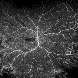
 Retinal Angiomas In VHL
Dec 24 2012 by Roy D. Brod, MD
Mid phase fluorescein angiogram of 16 year old male with recent diagnosis of Von Hippel-Lindau disease showing hyperfluorescent angioma in superior mid periphery OD.
Photographer: Julia Walker
Condition/keywords: hemangioma, Von Hippel-Lindau
 Loading…