Search results (333 results)
-
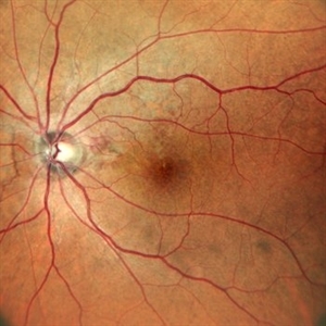
 Peripheral Exudative Hemorrhagic Chorioretinopathy
Oct 7 2020 by Olivia Rainey
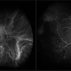
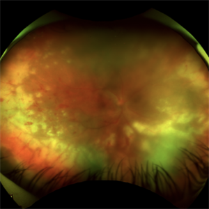
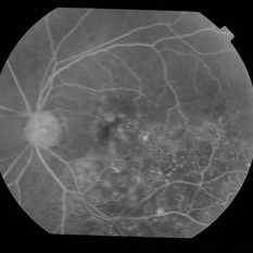
Fluorecein and ICG angiography of a 80-year-old male with peripheral exudative hemorrhagic chorioretinopathy affecting his right eye. Patient noted only mild floaters for a couple weeks OD on 10/5/2020. The physician strongly suspects that the lesion is subretinal blood (likely from PEHCR) rather than choroidal melanoma. There is blocking on FA and ICG with a definite lack of intrinsic vessels within the mass lesion. He will monitor closely, as the patient is monocular with a history of multiple surgeries (which the family believes PPV for "scar tissue") OS mostly in 2013. His family also reports remembering possibly being told there was a "small mass" in the left eye at one point in their surgical course. The physician believes that it's possible this was a bleed related to PEHCR as it typically exists as a bilateral condition.
Photographer: Olivia Rainey, OCT-C, COA
Imaging device: Optos California
Condition/keywords: fluorescein angiogram (FA), indocyanine green (ICG) angiography, monocular, Optos, peripheral exudative hemorrhagic chorioretinopathy (PEHCR), periphery, subretinal hemorrhage, ultra-wide field imaging
-
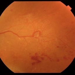 Venous Beading with Neovascularization
Jul 21 2025 by Moazzam Parvez
Fundus photograph of a 58 year old woman showing profuse venous beading and underlying neovascularization.
Photographer: Moazzam Parvez , Netralayam , Kolkata
Imaging device: Topcon Maestro 2
Condition/keywords: neovascularization (NV), periphery, Retina, venous beading
-
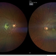 A rare case of a 45-year-old male with choroidal neovascular membrane in Familial Dominant Drusen (Doyne Honeycomb Drusen) in both eyes treated with intravitreal injections.
Nov 30 2022 by SHRADDHA ASHOK CHANDORKAR, DNB DO FVRS
A 45-year-old man presented with diminution of vision in both eyes with metamorphopsia, which was painless and gradually progressive in nature. BCVA at presentation were 6/40 and 6/36 for the right and left eye respectively. Anterior segment examination of both eyes was unremarkable. IOP were within normal limits. Fundus examination showed bilateral numerous yellowish white round closely spaced lesions extending radially from the vascular arcades till the periphery associated with an elevated grayish macular choroidal neovascular membrane (CNV) with multiple drusen in the macular area and posterior pole. Impression was Familial Dominant Drusen (Doyne Honeycomb Drusen) associated with CNVM, both eyes. Color fundus photograph and autofluorescence showed Familial Dominant Drusen with CNVM. Subsequently , the patient underwent periodic intravitreal injections of Ranibizumab in both eyes under guarded visual prognosis, for which he tolerated well.
Photographer: NATIONAL INSTITUTE OF OPHTHALMOLOGY, PUNE
Imaging device: ZEISS CLARUS
Condition/keywords: choroidal neovascular membrane (CNVM), Doyne's Honeycomb, FAMILIAL DOMINANT DRUSEN, IMIM (Online Mendelian Inheritance in Man), intravitreal injection, Malattia Leventinese
-
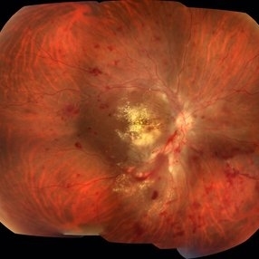 Acute Necrotizing Retinal Vasculitis as Onset of Systemic Lupus Erythematosus.
Sep 3 2016 by ADRIANO FERREIRA
A 28-year-old white man was referred to the rheumatology clinic with gradually and rapid deterioration of the vision (both eyes). In this picture, we can observe cotton wool spots in the papillomacular area and extensive hemorrhages in posterior polo and in the middle periphery. Hard exudates are present in macular area (macular edema)
Photographer: Claudio Zett Lobo
Imaging device: TRC50DXi TOPCON
Condition/keywords: systemic lupus erythematosus (SLE) vasculitis, vasculitis
-
 Acute Retinal Necrosis
Jul 3 2025 by Heitor Nogueira
Fundus photograph of an 53-year-old woman with patient who reported unilateral visual acuity loss for 10 days associated with ocular pain. She presented conjunctival hyperemia with temporal and nasal nodular scleritis, anterior chamber reaction 2+/4+, Koeppe nodules, granulomatous PKs, vitritis 2+/4+, multiple areas of vasculitis in arcades and periphery, associated with hemorrhages and necrotizing retinitis in temporal, inferior and nasal periphery. patient who reported unilateral visual acuity loss for 10 days associated with ocular pain. He presented conjunctival hyperemia with temporal and nasal nodular scleritis, anterior chamber reaction 2+/4+, Koeppe nodules, granulomatous PKs, vitreitis 2+/4+, multiple areas of vasculitis in the arcades and periphery, associated with hemorrhages and necrotizing retinitis in the temporal, inferior and nasal periphery. Positive serology for Herpes Virus.
Photographer: Heitor Nogueira, Penido Burnier Institute and CHOV, Campinas, São Paulo, Brazil
Imaging device: Optos Daytona
Condition/keywords: ARN complications, Herpes, progressive outer retinal necrosis (PORN)
-
 Acute Retinal Necrosis (ARN)
Jul 3 2025 by Heitor Nogueira
Fundus photograph of an 63-year-old woman who reported unilateral visual acuity loss for 10 days associated with ocular pain. He presented conjunctival hyperemia with temporal and nasal nodular scleritis, anterior chamber reaction 2+/4+, Koeppe nodules, granulomatous PKs, vitreitis 2+/4+, multiple areas of vasculitis in the arcades and periphery, associated with hemorrhages and necrotizing retinitis in the temporal, inferior and nasal periphery. Positive serology for Herpes Virus
Photographer: Heitor Nogueira, Penido Burnier Institute, Campinas, São Paulo, Brazil
Imaging device: Optos Daytona
Condition/keywords: ARN complications, Herpes, progressive outer retinal necrosis (PORN), Uveitis
-
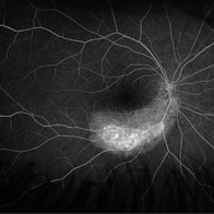 Acute Zonal Occult Outer Retinopathy (AZOOR) FA, Fluorescein Angiography, Peripheral Vasculitis
Jan 19 2022 by James B. Soque, CRA, OCT-C, COA, FOPS
Acute Zonal Occult Outer Retinopathy (AZOOR). Peripheral Vasculitis OD. Fluorescein angiography showing vasculitis in the far right periphery 8-10 o'clock. 46-year-old white male, VA CC 20/16, 20/12.5, has had recurrent vasculitis for 11 years. No treatment.
Photographer: James Soque, CRA, OCT-C, COA, FOPS, Island Retina, Shirley, NY
Imaging device: Optos California
Condition/keywords: acute zonal occult outer retinopathy (AZOOR), FA early phase, fluorescein angiogram (FA), Peripheral Vasculitis, ultra-wide field imaging
-
 Adult Coats' Disease
Aug 18 2015 by Mallika Goyal, MD
Left fundus of a 61-year-old non diabetic, non hypertensive lady complaining of vision deterioration for 1 year showing massive hard exudates at the macula. Fluorescein angiography revealed microvascular abnormalities over the posterior pole and temporal midperiphery and extensive capillary non-perfusion over the temporal retinal quadrants. OCT revealed macular edema. Fellow eye fundus and angiogram were normal.
Photographer: Mallika Goyal, MD, Apollo Health City, Jubilee Hills, Hyderabad
Condition/keywords: Coats' disease
-
 Adult Coats' Disease
Aug 18 2015 by Mallika Goyal, MD
Left fundus of a 61-year-old non diabetic, non hypertensive lady complaining of vision deterioration for 1 year showed massive hard exudates at the macula. Fluorescein angiography revealed microvascular abnormalities over the posterior pole and temporal midperiphery and extensive capillary non-perfusion over the temporal retinal quadrants. OCT revealed macular edema. Fellow eye fundus and angiogram were normal.
Photographer: Mallika Goyal, MD, Apollo Health City, Jubilee Hills, Hyderabad
Condition/keywords: Coats' disease
-
 Adult Coats' Disease
Aug 18 2015 by Mallika Goyal, MD
Fluorescein angiography of left eye of a lady with adult Coats' disease revealed microvascular abnormalities over the posterior pole and temporal midperiphery and extensive capillary non-perfusion over the temporal retinal quadrants. Other retinal areas had normal perfusion.
Photographer: Mallika Goyal, MD, Apollo Health City, Jubilee Hills, Hyderabad
Condition/keywords: Coats' disease
-
 Adult Coats' Disease
Aug 18 2015 by Mallika Goyal, MD
Fluorescein angiography of left eye of a lady with adult coats' disease revealed microvascular abnormalities over the posterior pole and temporal midperiphery and extensive capillary non-perfusion over the temporal retinal quadrants. Other retinal areas had normal perfusion.
Photographer: Mallika Goyal, MD, Apollo Health City, Jubilee Hills, Hyderabad
Condition/keywords: Coats' disease
-
 Adult Coats' Disease
Aug 18 2015 by Mallika Goyal, MD
Left fundus of a 61-year-old non diabetic, non hypertensive lady complaining of vision deterioration for 1 year showed massive hard exudates at the macula. Fluorescein angiography revealed microvascular abnormalities over the posterior pole and temporal midperiphery and extensive capillary non-perfusion over the temporal retinal quadrants. OCT revealed macular edema. Fellow eye fundus and angiogram were normal.
Photographer: Mallika Goyal, MD, Apollo Health City, Jubilee Hills, Hyderabad
Condition/keywords: Coats' disease
-
 Adult Coats' Disease
Aug 18 2015 by Mallika Goyal, MD
Left fundus of a 61-year-old non diabetic, non hypertensive lady complaining of vision deterioration for 1 year showed massive hard exudates at the macula. Fluorescein angiography revealed microvascular abnormalities over the posterior pole and temporal midperiphery and extensive capillary non-perfusion over the temporal retinal quadrants. OCT revealed macular edema. Fellow eye fundus and angiogram were normal.
Photographer: Mallika Goyal, MD, Apollo Health City, Jubilee Hills, Hyderabad
Condition/keywords: Coats' disease
-
 Advanced RP
Nov 5 2024 by rahul saradge
A man, 58, arrived complaining of BOV for both near and distance vision in both eyes, with a 6/9 BCVA in each eye. For a year, the patient had been taking medication for both diabetes and hypertension. In both eyes, the dilated ophthalmoscopic retina revealed waxy disc pallor paired with bony spicules in the mid-periphery. The patient was prescribed spectacles and given counseling regarding the nature of the illness.
Photographer: Lokesh Dukare ,Isha Netralaya Thane
Imaging device: optos
Condition/keywords: bone spicule, optic disc pallor, Optos, Retinitis Pigmentosa
-
 Angioid Streaks with Regressing CNV s/p AntiVEGF Injections (LE)
Sep 20 2024 by Anand Temkar
A 45 year old male came to our OPD with chief complaints of DOV in BE since 2 months and wavy vision in periphery. Patient was diagnosed with (BE) CNVM in a case of Angioid Streaks and has already received (BE) bevacizumab x 2.
Photographer: Dr.Anand Temkar- Retina Foundation, Ahmedabad
Imaging device: Mirante
Condition/keywords: Angioid Streaks, choroidal neovascularization (CNV), fundus autofluorescence (FAF)
-
 Angioid Streaks with Regressing CNV s/p AntiVEGF Injections (RE)
Sep 20 2024 by Anand Temkar
A 45 year old male came to our OPD with chief complaints of DOV in BE since 2 months and wavy vision in periphery. Patient was diagnosed with (BE) CNVM in a case of Angioid Streaks and has already received (BE) bevacizumab x 2.
Photographer: Dr.Anand Temkar- Retina Foundation, Ahmedabad
Imaging device: Mirante
Condition/keywords: Angioid Streaks, choroidal neovascularization (CNV), fundus autofluorescence (FAF)
-
 ARN (#1) Initial Photo
May 27 2019 by John S. King, MD
60-year-old African American female who had been treated for iridocyclitis for at least a week sent in for vitritis and a nasal fundus lesion. Complaints included redness, floaters, photophobia, and decreased vision. Husband had recent shingles. Acuity was 20/60-2 with IOP of 12, and small KP in Art's triangel, 1-2+ a/c cell, 2-3+ ant vit cell, diffuse arteriolar sheathing, multiple areas of retinal whitening in periphery and mid-periphery (see Photo #1). PCR of a/c was performed, and intravitreal GCV administered, and VACV 2g qid and ASA started.... PCR positive for HZV, pred taper was started two days after presentation as the infection had begun to stablize..... Five days from presentation the vision was 20/60, inflammation and areas of retinal whitening had improved (see Photo #2).... One week later acuity was 20/30, the a/c was quiet and KP resolved; ant vitreous cell decreased; and there was further improvement in retinal appearance without any signs of retinal holes or detachment; she is now on low dose maint VACV (see photo#3)
Photographer: Maysee Yang
Imaging device: Optos CA
Condition/keywords: acute retinal necrosis, Herpes zoster
 Loading…