Search results (333 results)
-
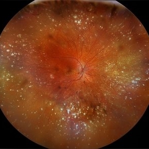
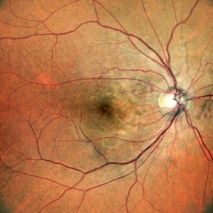
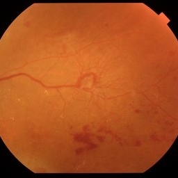 Venous Beading with Neovascularization
Jul 21 2025 by Moazzam Parvez
Fundus photograph of a 58 year old woman showing profuse venous beading and underlying neovascularization.
Photographer: Moazzam Parvez , Netralayam , Kolkata
Imaging device: Topcon Maestro 2
Condition/keywords: neovascularization (NV), periphery, Retina, venous beading
-
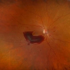
 Large Subhyaloid Hemorrhage
Jul 11 2025 by Jessilla Phou
This is a fundus photograph depicting a large subhyaloid hemorrhage in the mid periphery of the left eye. The patient, a 53-year-old female, presented with a sudden onset of floaters, headache, and blurred vision. The image also demonstrates associated optic disc hemorrhage, vitreous hemorrhage, retinal hemorrhage, and venous tortuosity. Despite the extensive workup performed and the severity of the hemorrhage, no underlying cause was determined.
Photographer: Jessilla Phou
Imaging device: Optos California
Condition/keywords: fundus photograph, optic disc hemorrhage, retinal hemorrhage, venous tortuosity, vitreous hemorrhage
-
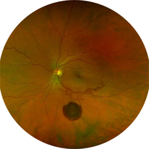
 Pseudoduplication of the Optic Disc
Jul 9 2025 by Hrishikesh Naik, MS
A peripapillary colobomatous pseudo-duplication of the optic disc as seen in an asymptomatic 23 year old female with myopia referred for routine retinal periphery screening. Rest retinal exam was normal. Duplication of the optic disc can be classified as either true duplication or pseudoduplication, both of which are rare clinical conditions. Pseudodoubling of the optic disc is commonly caused by optic disc or peripapillary colobomas, characterized by a circumscribed, disc-like lesion with radiating vessels but only one normal optic nerve. A few cases have involved pathological myopia, moderate myopia, proliferative diabetic retinopathy and CHARGE syndrome. The lesion is often found inferior to the normal optic disc. The patient was advised regular follow ups.
Photographer: Hrishikesh Naik
Imaging device: Optos Daytona
Condition/keywords: Coloboma, Pseudoduplication of optic disc
-
 Acute Retinal Necrosis (ARN)
Jul 3 2025 by Heitor Nogueira
Fundus photograph of an 63-year-old woman who reported unilateral visual acuity loss for 10 days associated with ocular pain. He presented conjunctival hyperemia with temporal and nasal nodular scleritis, anterior chamber reaction 2+/4+, Koeppe nodules, granulomatous PKs, vitreitis 2+/4+, multiple areas of vasculitis in the arcades and periphery, associated with hemorrhages and necrotizing retinitis in the temporal, inferior and nasal periphery. Positive serology for Herpes Virus
Photographer: Heitor Nogueira, Penido Burnier Institute, Campinas, São Paulo, Brazil
Imaging device: Optos Daytona
Condition/keywords: ARN complications, Herpes, progressive outer retinal necrosis (PORN), Uveitis
-
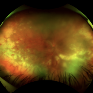
 Acute Retinal Necrosis
Jul 3 2025 by Heitor Nogueira
Fundus photograph of an 53-year-old woman with patient who reported unilateral visual acuity loss for 10 days associated with ocular pain. She presented conjunctival hyperemia with temporal and nasal nodular scleritis, anterior chamber reaction 2+/4+, Koeppe nodules, granulomatous PKs, vitritis 2+/4+, multiple areas of vasculitis in arcades and periphery, associated with hemorrhages and necrotizing retinitis in temporal, inferior and nasal periphery. patient who reported unilateral visual acuity loss for 10 days associated with ocular pain. He presented conjunctival hyperemia with temporal and nasal nodular scleritis, anterior chamber reaction 2+/4+, Koeppe nodules, granulomatous PKs, vitreitis 2+/4+, multiple areas of vasculitis in the arcades and periphery, associated with hemorrhages and necrotizing retinitis in the temporal, inferior and nasal periphery. Positive serology for Herpes Virus.
Photographer: Heitor Nogueira, Penido Burnier Institute and CHOV, Campinas, São Paulo, Brazil
Imaging device: Optos Daytona
Condition/keywords: ARN complications, Herpes, progressive outer retinal necrosis (PORN)
-
 VKH Syndrome
Jun 12 2025 by Virginia Gebhart
22 year old male with VKH Syndrome. Pt has been experiencing severe headaches, distorted vision, hearing loss, weakness, and a large white patch of hair. Significant cell in AC and vitreous, multiple punched-out CR scars in periphery. Referred to rheumatology for possible immunomodulatory treatment
Photographer: Virginia Gebhart, Retina Consultants of Carolina
Imaging device: Optos California
Condition/keywords: montage, multifocal choroiditis, panuveitis, Vogt-Koyanagi-Harada
-
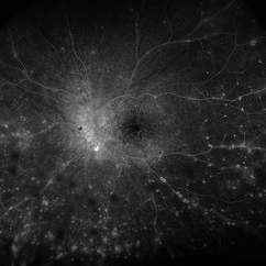 VKH Syndrome
Jun 12 2025 by Virginia Gebhart
Fluorescein angiogram of 22 year old male with VKH syndrome. Significant cell in AC and vitreous, multiple punched-out CR scars in periphery, mild vascular leakage. Pt referred to rheumatology for immunomodulatory treatment.
Photographer: Virginia Gebhart, Retina Consultants of Carolina
Imaging device: Optos California
Condition/keywords: FA, fluorescein angiogram (FA), multifocal choroiditis, panuveitis, VKH, Vogt-Koyanagi-Harada
-
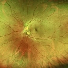
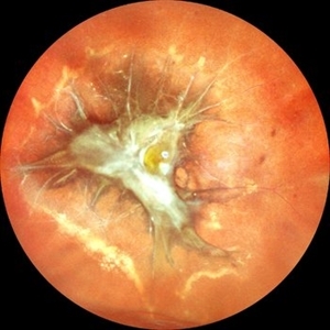 Traction in Proliferative Diabetic Retinopathy
Jun 9 2025 by Malvika Singh
Fundus photograph of a 44 year old with uncontrolled diabetes showing fibrovascular proliferation and traction with details of disc and macula obscured with sclerosed vessels in the periphery.
Photographer: Dr Malvika Singh, Retina Foundation, Ahmedabad, India
Imaging device: Mirante SLO/OCT
Condition/keywords: proliferative diabetic retinopathy (PDR), TRACTION
-
 Macular Schisis with RD
Apr 4 2025 by Tejaswita Verma
Fundus photo of a 29 year-old male with RRD, breaks in periphery, macular schisis . Vision CF2.5 mts
Photographer: DR. TEJASWITA VERMA
Imaging device: MIRANTE
Condition/keywords: macular schisis, retinal detachment
-
 Vasoproliferative Tumor
Mar 25 2025 by Gustavo Uriel Fonseca Aguirre
Patient diagnosed with pars planitis and a history of phacovitrectomy. Longitudinal B-scan section showing a very pronounced, homogeneous tumor lesion in the periphery. The A-scan revealed high average reflectivity with an irregular internal structure.
Photographer: Gustavo U. Fonseca Aguirre, Hospital Oftalmológico de la Luz, Ciudad de México
Condition/keywords: pars planitis, Vasoproliferative Tumor
-
 Proliferative Diabetic Retinopathy S/P Pan Retinal Photocoagulation
Mar 4 2025 by Prithvi Chandrakanth
A 52-year-old female patient presented with complaints of diminishing vision, compounded by uncontrolled diabetes mellitus. Her Fundus examination revealed proliferative diabetic retinopathy, characterized by neovascularization of the disc and elsewhere, and sclerosed vessels. To address this, Pan Retinal Photocoagulation was performed, and the condition stabilized, halting the progression of the disease.
Photographer: DR PRITHVI CHANDRAKANTH, DR CHANDRAKANTH NETHRALAYA, KOZHIKODE, KERALA, INDIA
Imaging device: EIDON
Condition/keywords: Diabetic Retinopathy, Neovascularisation at the Disc (NVD), neovascularization of the disc (NVD), NVD, pan-retinal photocoagulation (PRP), PDR, PDR with NVE (periphery), PRP
-
 Asteroid Hyalosis in Retinitis Pigmentosa
Dec 9 2024 by Mauricio Bayram-Suverza, MD
A 54 year-old male patient presented with asteroid hyalosis. Retinal examination revealed the presence of bone spicules, primarily located in the mid-periphery. Genetic testing identified a pathogenic variant in the RHO gene.
Photographer: Mauricio Bayram-Suverza, Casey Eye Institute, OHSU.
Imaging device: Optos California
Condition/keywords: Asteroid hyalosis, retinal dystrophy, Retinitis Pigmentosa, vitreous
-
 Advanced RP
Nov 5 2024 by rahul saradge
A man, 58, arrived complaining of BOV for both near and distance vision in both eyes, with a 6/9 BCVA in each eye. For a year, the patient had been taking medication for both diabetes and hypertension. In both eyes, the dilated ophthalmoscopic retina revealed waxy disc pallor paired with bony spicules in the mid-periphery. The patient was prescribed spectacles and given counseling regarding the nature of the illness.
Photographer: Lokesh Dukare ,Isha Netralaya Thane
Imaging device: optos
Condition/keywords: bone spicule, optic disc pallor, Optos, Retinitis Pigmentosa
-
 Torpedo Retinopathy
Oct 31 2024 by AVIK DEY SARKAR, MS, FVRS, FAICO(VR)
This is a 42 year old male with known history of diabetes mellitus for past 10 years. Patient presented with complains regarding presbyopia. On dilated fundoscopy, along with dot and blot hemorrhages, in the infero-temporal near-periphery outside the vascular arcade a hypopigmented torpedo-shaped lesion was noted. On OCT, outer retinal attenuation with sublesional choriocappilaris layer thinning was noted. The lesion is diagnosed as torpedo retinopathy. Torpedo maculopathy is rare in clinical practice and usually is found at the margin of temporal arcade over "Temporal Bulge". But this lesion is seen well away from the posterior pole. This case indicates the necessity of substituting the terminology "Torpedo Maculopathy" with "Torpedo Retinopathy" as mentioned earlier in ophthalmic literature.
Photographer: Dr. Avik Dey Sarkar, MBBS, MS, FVRS, FAICO, Consultant, Department of Vitreoretinal Services, Aravind Eye Hospital, Madurai, India
Imaging device: Wide angled Fundus imaging with Clarus 300
Condition/keywords: torpedo maculopathy, torpedo Retinopathy
-
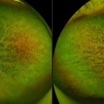
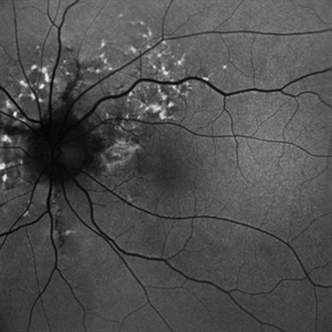 Fundus Autofluorescence Showing Angioid Streaks with Regressing CNV s/p AntiVEGF Injections (LE)
Sep 20 2024 by Anand Temkar
A 45 year old male came to our OPD with chief complaints of DOV in BE since 2 months and wavy vision in periphery. Patient was diagnosed with (BE) CNVM in a case of Angioid Streaks and has already received (BE) bevacizumab x 2.
Photographer: Dr.Anand Temkar- Retina Foundation, Ahmedabad
Imaging device: Mirante
Condition/keywords: Angioid Streaks, choroidal neovascularization (CNV), fundus autofluorescence (FAF)
-
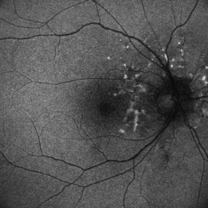 Fundus Autofluorescence Showing Angioid Streaks with Regressing CNV s/p AntiVEGF Injections (RE)
Sep 20 2024 by Anand Temkar
A 45 year old male came to our OPD with chief complaints of DOV in BE since 2 months and wavy vision in periphery. Patient was diagnosed with (BE) CNVM in a case of Angioid Streaks and has already received (BE) bevacizumab x 2.
Photographer: Dr.Anand Temkar- Retina Foundation, Ahmedabad
Imaging device: Mirante
Condition/keywords: Angioid Streaks, choroidal neovascularization (CNV), fundus autofluorescence (FAF)
-
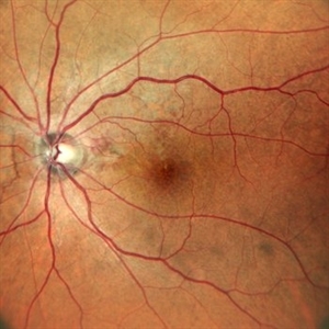 Angioid Streaks with Regressing CNV s/p AntiVEGF Injections (LE)
Sep 20 2024 by Anand Temkar
A 45 year old male came to our OPD with chief complaints of DOV in BE since 2 months and wavy vision in periphery. Patient was diagnosed with (BE) CNVM in a case of Angioid Streaks and has already received (BE) bevacizumab x 2.
Photographer: Dr.Anand Temkar- Retina Foundation, Ahmedabad
Imaging device: Mirante
Condition/keywords: Angioid Streaks, choroidal neovascularization (CNV), fundus autofluorescence (FAF)
-
 Angioid Streaks with Regressing CNV s/p AntiVEGF Injections (RE)
Sep 20 2024 by Anand Temkar
A 45 year old male came to our OPD with chief complaints of DOV in BE since 2 months and wavy vision in periphery. Patient was diagnosed with (BE) CNVM in a case of Angioid Streaks and has already received (BE) bevacizumab x 2.
Photographer: Dr.Anand Temkar- Retina Foundation, Ahmedabad
Imaging device: Mirante
Condition/keywords: Angioid Streaks, choroidal neovascularization (CNV), fundus autofluorescence (FAF)
-
 OCT in Case of Macular Coloboma (LE)
Sep 18 2024 by Anand Temkar
A 24 year old male came with chief complaint of diminution of vision in both eyes since childhood. Vision in both eyes was 6/24. IOP in RE was 12 and LE was 14 mm of Hg. On fundus examination periphery was within normal limits and central fundus revealed this picture. The serology testing such as serum IgM, IgG for toxoplasma and cytomegalovirus was negative. I have also uploaded LE color photo and BE OCT of this patient.
Photographer: Dr.Anand Temkar- Retina Foundation, Ahmedabad
Imaging device: Mirante
Condition/keywords: Coloboma
-
 OCT in Case of Macular Coloboma (RE)
Sep 18 2024 by Anand Temkar
A 24 year old male came with chief complaint of diminution of vision in both eyes since childhood. Vision in both eyes was 6/24. IOP in RE was 12 and LE was 14 mm of Hg. On fundus examination periphery was within normal limits and central fundus revealed this picture. The serology testing such as serum IgM, IgG for toxoplasma and cytomegalovirus was negative. I have also uploaded LE color photo and BE OCT of this patient.
Photographer: Dr.Anand Temkar- Retina Foundation, Ahmedabad
Imaging device: Mirante
Condition/keywords: coloboma
-
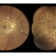
 Macular Coloboma (LE)
Sep 18 2024 by Anand Temkar
A 24 year old male came with chief complaint of diminution of vision in both eyes since childhood. Vision in both eyes was 6/24. IOP in RE was 12 and LE was 14 mm of Hg. On fundus examination periphery was within normal limits and central fundus revealed this picture. The serology testing such as serum IgM, IgG for toxoplasma and cytomegalovirus was negative. I have also uploaded LE color photo and BE OCT of this patient.
Photographer: Dr.Anand Temkar- Retina Foundation, Ahmedabad
Imaging device: Mirante
Condition/keywords: macular coloboma
-
 Macular Coloboma (RE)
Sep 18 2024 by Anand Temkar
A 24 year old male came with chief complaint of diminution of vision in both eyes since childhood. Vision in both eyes was 6/24. IOP in RE was 12 and LE was 14 mm of Hg. On fundus examination periphery was within normal limits and central fundus revealed this picture. The serology testing such as serum IgM, IgG for toxoplasma and cytomegalovirus was negative. I have also uploaded LE color photo and BE OCT of this patient.
Photographer: Dr.Anand Temkar- Retina Foundation, Ahmedabad
Imaging device: Mirante
Condition/keywords: coloboma of macula
-
 Pre-Retinal Hemorrhage
Aug 22 2024 by Virginia Gebhart
51 year old female with moderate proliferative diabetic retinopathy, DME, as well as pre-retinal hemorrhage and likely NVE. Pt given Avastin in office and will return for PRP.
Photographer: Virginia Gebhart
Imaging device: Optos California
Condition/keywords: diabetic macular edema, macular edema, PDR with NVE (periphery), pre-retinal hemorrhage, proliferative diabetic retinopathy (PDR)
-
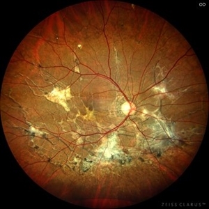 Pseudoxanthoma Elasticum Associated Angioid Streaks
Aug 18 2024 by KANWALJEET HARJOT MADAN, M.S. (Ophthalmology); FAICO (Vitreous - Retina)
This is fundus photograph of a young 31 years male patient depicting Angioid streaks emanating from optic nerve towards the periphery and subretinal fibrosis. There is peau de orange appearance temporal to fovea with Salmon Spots in periphery. He was diagnosed to have Pseudoxanthoma Elasticum.
Photographer: Dr. Kanwaljeet Harjot Madan, M.S. (Ophthalmologist) Fellow in Vitrous & Retina. Thind Eye Hospital, Jalandhar City. Punjab. India
Imaging device: Zeiss Clarus
Condition/keywords: Angioid Streaks, fundus photograph, pseudoxanthoma elasticum (PXE)
-
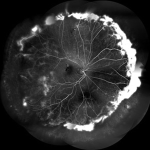 FFA in a Case of Retinoschisis With Fluid
Jul 28 2024 by Prashant K Bawankule, M.S.
Young male of 22 years presented with DOV. Examination showed retinoschisis with fluid in periphery. FFA showed massive leakage in the periphery
Photographer: Prashant Bawankule, Sarakshi Netralaya, Nagpur, Maharashtra , India
Imaging device: Mirante ( by Nidek)
Condition/keywords: retinoschisis
 Loading…