Search results (333 results)
-
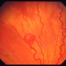
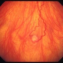
 Familial Dominant Drusen
Nov 22 2015 by Mallika Goyal, MD
Bilateral drusen over the entire retinal mid-periphery and periphery of a 29-year-old male with no visual complaints. Macular centre is normal though there are some drusen in the temporal macula.
Photographer: Mallika Goyal, MD, Apollo Health City, Jubilee Hills, Hyderabad, India
Condition/keywords: familial drusen
-
 Snail Track Peripheral Retinal Degeneration
Apr 29 2022 by Otakar Dušek, M.D. Ph.D.
Colour fundus photograph of 22-year-old woman with incidentally found snail track retinal degeneration in the superior temporal periphery of the retina of the right eye.
Photographer: Otakar Dušek, Charles University, Prague
Imaging device: Zeiss Clarus
Condition/keywords: peripheral retinal degeneration
-
 Commotio retinae
Jan 11 2013 by Alex P. Hunyor, MD
Commotio retinae in the temporal mid periphery in an eye which sustained blunt trauma.
Condition/keywords: Berlin's edema, commotio retinae
-
 Retinal capillary hemangiomas 3
Jan 11 2013 by Alex P. Hunyor, MD
Retinal capillary haemangiomas, left superior periphery, in a 20 year old female with von Hippel-Lindau disease.
Condition/keywords: hemangioma, retinal capillary hemangioma, Von Hippel-Lindau
-
 Familial Dominant Drusen
Nov 22 2015 by Mallika Goyal, MD
Bilateral drusen over the entire retinal mid-periphery and periphery of a 29-year-old male with no visual complaints. Macular centre is normal though there are some drusen in the temporal macula.
Photographer: Mallika Goyal, MD, Apollo Health City, Jubilee Hills, Hyderabad, India
Condition/keywords: familial drusen
-
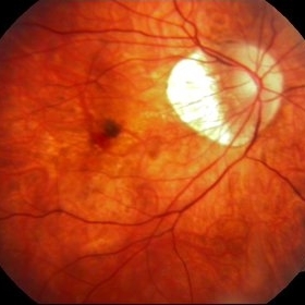 Myopic Choroidal Neovascular Membrane
Mar 25 2013 by Ratimir Lazic, MD, PhD
Color fundus photography of a 33-year-old female. In macular area subretinal hemorrhage can be seen. Area of atrophy temporal from PNO. Myopic changes of posterior pole and mid periphery can be noticed. The patient has been treated with 2 consecutive ranibizumab intravitreal injections. BCVA at baseline was 0,05 (Snellen lines) and 0,3 (Snellen lines) 2 months after.
Photographer: Marko Lukic, MD
Imaging device: Zeis Visucam Lite 2
Condition/keywords: high myopia, myopic choroidal neovascularization (CNV), ranibizumab
-
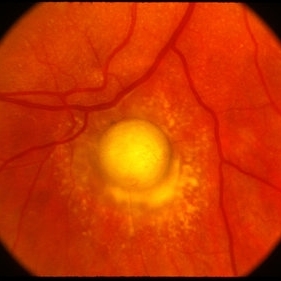 Toxocara Granuloma
Feb 25 2013 by Henry J. Kaplan, MD
Toxocara granuloma in the midperiphery of the retina.
Condition/keywords: ocular toxoplasmosis, toxocara granuloma, toxocariasis
-
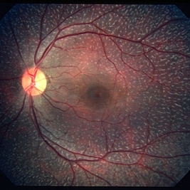 Fundus Albipunctatus
Mar 29 2013 by Henry J. Kaplan, MD
Fundus albipunctatus (one of the stationary night blindness syndromes with multiple white dots in the periphery and normal optic disc and vessels).
Condition/keywords: fundus albipunctatus
-
 Stargardts Disease in FAF
Sep 14 2012 by Michael P. Kelly, FOPS
This is a scanning laser ophthalmoscopic FAF image of a patient with Stargardts Disease captured with a Heidelberg Spectralis imaging unit. Note, besides the obvious hyper-autofluorescent areas centrally, the much smaller, and in greater number, pinpoints of hyper-autofluorescence extending from the vascular arcades into the mid-periphery.
Photographer: Michael P. Kelly, FOPS, Director, Duke Eye Center Labs, Duke Universtiy Hospital
Imaging device: Heidelberg Spectralis
Condition/keywords: fundus autofluorescence (FAF), Stargardt disease
-
 Retinal capillary hemangioma 2
Jan 11 2013 by Alex P. Hunyor, MD
Retinal capillary haemangioma, right inferior periphery, in a 20-year-old female with von Hippel-Lindau disease.
Condition/keywords: hemangioma, retinal capillary hemangioma, Von Hippel-Lindau
-
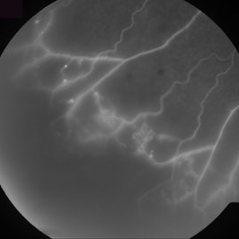
 Ischemic BRVO with neovascularization
Aug 23 2012 by Gerardo Garcia-Aguirre, MD
Fluorescein angiogram of the temporal periphery showing wide areas of capillary nonperfusion and leakage secondary to neovascularization.
Photographer: Noemí Hernández, Asociación para Evitar la Ceguera en México
Condition/keywords: branch retinal vein occlusion (BRVO), capillary nonperfusion, neovascularization (NV)
-
 Retinal Angiomas In VHL
Dec 24 2012 by Roy D. Brod, MD
Fundus photograph of 16 year old male with recent diagnosis of Von Hippel-Lindau disease showing typical appearance of a retinal angioma in superior mid periphery OD. Note unrelated choroidal nevus above superior arcade.
Photographer: Julia Walker
Condition/keywords: hemangioma, Von Hippel-Lindau
-
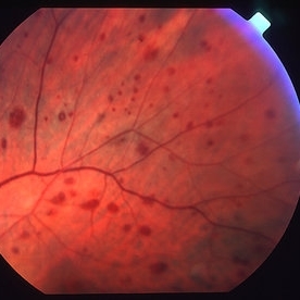 Ocular ischaemic syndrome colour 2
Jan 11 2013 by Alex P. Hunyor, MD
Ocular ischaemic syndrome, left eye - color image, superotemporal midperiphery. Note: dilated but not tortuous veins, attenuated arteries, and multiple intraretinal haemorrhages.
Condition/keywords: ocular ischemic syndrome
-
---thumb.jpg/image-square;max$300,300.ImageHandler) Multifocal Choroiditis
Multifocal Choroiditis
Feb 26 2013 by Henry J. Kaplan, MD
Multifocal choroiditis, MFC, inactive scars in the periphery.
Condition/keywords: multifocal choroiditis
-
 Atrophic Scar
Oct 16 2012 by Ratimir Lazic, MD, PhD
Color fundus image of a 76-year-old female. In this color image the atrophic scar in large macular area and mild periphery can be seen. BCVA on that eye is CF on 1m.
Photographer: Marko Lukic, MD
Imaging device: Zeis Visucam Lite 2
Condition/keywords: atrophic scar, retinal pigment epithelium
-
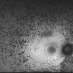 Retinitis Pigmentosa - Autofluorescence OD
Jun 18 2018 by Hosam Attia, MD
Ultra-wide fundus auto-fluorescence photograph of a 38-year-old African, American female with degenerative myopia, unilateral RP variant, depicting extensive mid-peripheral bone spicules hypo-autofluorescence, extending further into the periphery w/ relative sparing of the macula OD VF 30-V showed severe peripheral constriction OD, enlarged BS OS and OCT showed severe ellipsoid zone degeneration with saucerization and cystoid macular degeneration with no obvious late macular leakage on FA (Both, not shown)
Imaging device: Optos California
Condition/keywords: bone spicule, peripheral bone spicules, retinitis pigmentosa
-
 Multiple Evanescent White Dot Syndrome (MEWDS)
Oct 20 2012 by Hyung-Woo Kwak, MD
Numerous small deep ill-defined, grey-white dot were seen at the posterior pole and mid-periphery. Some lesions showed mild hyperfluorescence in autofluorescence (AF) but were of limited diagnostic value. ICG showed more numerous hypofluorescent spots than are apparent clinically or on AF/FA
Condition/keywords: hypofluorescent spots, multiple evanescent white dot syndrome (MEWDS)
-
Stargardts Disease in Fundus Autofluorescence
Sep 12 2012 by Michael P. Kelly, FOPS
Fundus autofluorescence of a patient with Stargardts disease. Note the central area of hypo-autofluorescence indicating atrophy surrounded by smaller areas of hyper-autofluorescence. Note also the much smaller, and in greater number, pinpoints of hyper-autofluorescence extending from the vascular arcades into the mid-periphery.
Photographer: Michael P. Kelly, FOPS, Director, Duke Eye Labs, Duke University Hospital, Duke Eye Center
Imaging device: Heidelberg Spectralis
Condition/keywords: fundus autofluorescence (FAF), Stargardt disease
-
 Retinal Angiomas In VHL
Dec 24 2012 by Roy D. Brod, MD
Fundus photograph of 16 year old male with recent diagnosis of Von Hippel-Lindau disease showing 2 retinal angiomas in inferior mid periphery OD.
Photographer: Julia Walker
Condition/keywords: hemangioma, Von Hippel-Lindau
-
 Ischemic BRVO
Aug 23 2012 by Gerardo Garcia-Aguirre, MD
Fluorescein angiogram of the inferotemporal periphery showing wide areas of capillary nonperfusion.
Photographer: Noemí Hernández, Asociación para Evitar la Ceguera en México
Condition/keywords: branch retinal vein occlusion (BRVO), capillary nonperfusion
-
 Familial Dominant Drusen
Nov 22 2015 by Mallika Goyal, MD
Bilateral drusen over the entire retinal mid-periphery and periphery of a 29-year-old male with no visual complaints. Macular centre is normal though there are some drusen in the temporal macula.
Photographer: Mallika Goyal, MD, Apollo Health City, Jubilee Hills, Hyderabad, India
Condition/keywords: familial drusen
-
 Multiple Evanescent White Dot Syndrome (MEWDS)
Oct 20 2012 by Hyung-Woo Kwak, MD
Numerous small deep ill-defined, grey-white dot were seen at the posterior pole and mid-periphery. Some lesions showed mild hyperfluorescence in autofluorescence (AF) but were of limited diagnostic value. ICG showed more numerous hypofluorescent spots than are apparent clinically or on AF/FA
Condition/keywords: hypofluorescent spots, multiple evanescent white dot syndrome (MEWDS)
-
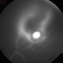 Toxoplasma Gondii Chorioretinitis, Fluorescein Angiogram
Aug 23 2012 by Gerardo Garcia-Aguirre, MD
Fluorescein angiogram of the superior periphery showing a highly hyperfluorescent lesion adjacent to a hypofluorescent lesion, surrounded by a hyperfluorescent halo.
Photographer: Noemí Hernández, Asociación para Evitar la Ceguera en México
Imaging device: Zeiss FF4
Condition/keywords: chorioretinal inflammations, toxoplasmosis
-
 Retinal Angiomas In VHL
Dec 24 2012 by Roy D. Brod, MD
Mid phase fluorescein angiogram of 16 year old male with recent diagnosis of Von Hippel-Lindau disease showing hyperfluorescent angioma in superior mid periphery OD.
Photographer: Julia Walker
Condition/keywords: hemangioma, Von Hippel-Lindau
-
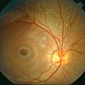 Niemann Pick Disease Type B
Aug 6 2013 by Hamid Ahmadieh, MD
Color fundus photograph of the right eye of a patient with Niemann Pick Type B with typical macular halo and confluent deposits in mid-periphery.
Photographer: Ali Mohammad-Rabie, Ophthalmic Research Center, Labbafinejad Medical Center, Tehran
Condition/keywords: fleck retinopathy, macular halo
 Loading…