File number: 20572
Comments
-
 Avris Romario Diparaja Siahaan (September 11 2015)
Thank you for your kindly comment Sir....
I will tell it to my friend Harni
:-) -
 James B. Soque, CRA, OCT-C, COA, FOPS (September 10 2015)
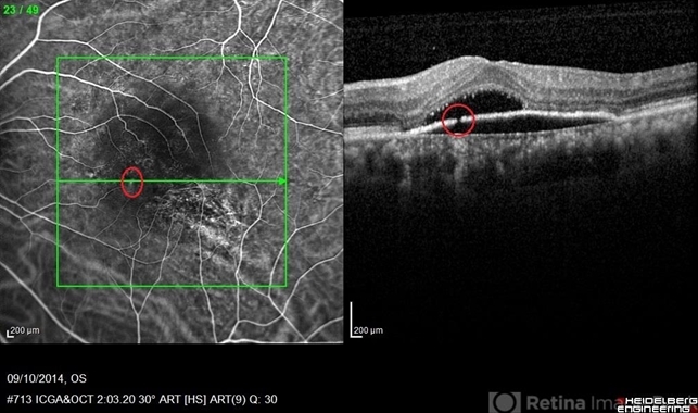
This is a superb example of imaging an RPE detachment, and documenting a break in Bruch's membrane causeing the formation of the serous detachment in this patients left eye. A rare use of the utility of simultaneous ICG Angiography and OCT using a Heigelberg Spectrlis device. You do our profession justice Harni, well done!
Sign in to comment.
 Initializing download.
-
 By Avris Romario Diparaja Siahaan
- Uploaded on Oct 17, 2014.
- Last modified by Caroline Bozell on Oct 20, 2014.
- Rating
- Appears in
- Discontinuity RPE
- Condition/keywords
- optical coherence tomography (OCT), retinal pigment epithelium, indocyanine green (ICG) angiography
- Photographer
- Harni Christine Damanik, Klinik Mata Nusantara
- Imaging device
-
Scanning laser ophthalmoscope
Heidelberg Spectralis - Description
- A simultan ICG angiography + OCT of 56-year-old man that shows a image of discontinuity retinal pigment ephitelial.
")


")


