Search results (76 results)
-
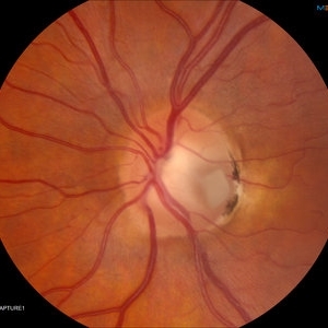
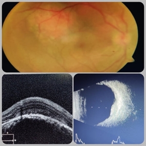
 Choroidal Fracture
Oct 27 2024 by César Adrián Gómez Valdivia, MD
Fundus photograph of a traumatic choroidal fracture & extra-macular sub-retinal hemorrhage.
Photographer: @eyemissu2
Imaging device: TOPCON TRC-50DX
Condition/keywords: Choroidal Fracture
-
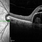
 Central Serous Retinopathy
Mar 19 2024 by Corey Grant
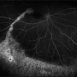
Ultra Wide-Field Fundus Autofluorescence Imaging of a 37 year old female with Central Serous Retinopathy affecting her right eye. Patient Visual Acuity was 20/20 in both eyes. Patient reported black spots in her vision onset three years ago, with associating flashes of light. Patient reports history of cortisone back injections a few years ago and denies Flonase use. The physician stated that there is hyperautofluorescence in the area of gutter of Sub-Retinal Fluid which likely happened from CSR.
Photographer: Corey Grant, OSC
Imaging device: OPTOS CALIFORNIA RGB
Condition/keywords: Central Serous Chorioretinopathy (CSR), central serous retinopathy (CSR), fundus autofluorescence (FAF), Guttering, hyperautofluorescence, inferior retina, OPTOS, Retina, Right Eye, subretinal fluid, ULTRA WIDE FIELD
-
 Ocular Toxocariasis slide 3
Oct 22 2012 by Ronald C. Gentile, MD
The sub-retinal scarred granuloma was white in color and elevated. It had pigment speckling around it. Serum testing was positive for past exposure to Toxocara canis.
Photographer: The New York Eye & Ear Infirmary Department of Medical Imaging
Condition/keywords: toxocariasis
-
 Optic Nerve Pit With Sub-Retinal Fluid
Sep 17 2015 by Jason S. Calhoun
Young female with blurred vision in the left eye. Fundus photograph shows optic nerve pit adjacent to the macula where there is sub retinal fluid visible.
Photographer: Jason Calhoun, Mayo Clinic, Department of Ophthalmology
Imaging device: TOPCON-TRC50EX
Condition/keywords: congenital optic nerve pit
-
 Pigment Epithelium Detachment, Secondary to AMD
Mar 17 2023 by Ceara Donovan
Optical coherence tomography of a 76 year old woman with a Pigment Epithelium Detachment, Secondary to AMD affecting her right eye. Patient had no significant response to Avastin, Eylea, Lucentis 0.5, or Vabysmo and was switched to Beovu. Following Beovu intravitreal injection her edema improved on OCT. Patient's vision was sc20/200+1 at the time the image was taken.
Photographer: Ceara Donovan
Imaging device: Heidelberg Spectralis
Condition/keywords: exudative age-related macular degeneration, heidelberg spectralis, macular degeneration, optical coherence tomography (OCT), pigment epithelial detachment (PED), Sub-retinal fluid
-
 Subretinal Bleed
Jul 12 2022 by Akansha Sharma
73 year old diabetic and hypertensive female presented with sub-retinal hemorrhage for which she was operated with pars-plana vitrectomy with intra-vitreal anti-VEGF
Photographer: Dr. Akansha Sharma-Retina Foundation, Ahmedabad
Condition/keywords: subretinal hemorrhage, subretinal blood
-
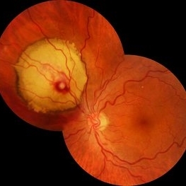
 Multiple Retinal Lesions Secondary to Blunt Trauma
Jun 19 2018 by Somnath Chakraborty, MD
A montage of the right eye of a 15-year-old boy, who was struck by a football. The image shows multiple choroidal ruptures in the macular area, with sub-retinal blood and multiple, large retinal tears temporally. There is also an area of juxtapapillary, pigmentary changes.
Photographer: Saptarshi Mehta, Retina Institute of Bengal
Condition/keywords: blunt trauma, choroidal rupture, giant retinal tear, subretinal hemorrhage
-
 Retinal Arterial Macroaneurysm
Sep 18 2018 by Somnath Chakraborty, MD
Left eye fundus photo of a 72-year-old hypertensive, female with a hemorrhagic retinal arterial macroaneurysm with sub-retinal blood.
Photographer: Pulok Chandra Roy, Retina Institute of Bengal
Condition/keywords: retinal arterial macroaneurysm
-
 24 Hours Post Scleral Wound Closure+ Scleral Buckle+25 g Vitrectomy+Silicon Oil
Jan 23 2015 by Carlos Quezada-Ruiz, MD, FASRS
24 hours post op fundus photograph of a 43-year-old man who had perforating injury to the right eye with a small piece of plastic while he was hammering. OD LP, subconjunctival hemorrhage, clear cornea, hyphema, irido and ciclodyalisis as well as a luxated lens with traumatic cataract and a dense vitreous hemorrhage. B-US showed rhegmatogenous retinal detachment with a tear and a big inferior hemorrhagic choroidal detachment. 360 peritomy revealed 2-entry scleral wounds were found in zone II (M V and M VI) and closure was performed. 25 G PPV was performed with the infusion canal placed in the AC through the limbus. Lensectomy and removal of a dense recent vitreous hemorrhage revealed a white detached retina with an exit wound through the temporal inferior segment of the optic nerve with a nasal GRT and sub retinal hemorrhage as well as temporal inferior choroidal, PVD was induced and PFOs helped stabilizing the retina while vitrectomy and sub-retinal hemorrhage was removed through the GRT. Fluid air exchange was made and 360 endolaser over the buckle indentation was done and silicon oil was used as endotamponade. This picture was taken 24 hrs after the surgery.
Photographer: Lilibeth Rodriguez, Instituto de la Visión. Torreon, Mexico.
Condition/keywords: central retinal artery occlusion (CRAO), giant retinal tear, trauma
-
 choroidal lymphoma
Nov 25 2012 by Mallika Goyal, MD
Left eye of a 60-year-old lady shows multiple sub-retinal yellowish masses of choroidal lymphoma. Radiotherapy resulted in complete regression with recurrence after 10 months.
Photographer: Mallika Goyal, MD, Apollo Health City, Hyderabad, India
Condition/keywords: lymphoma
-
 Chronic Central Serous Chorioretinopathy
Mar 29 2019 by Nichole Lewis
54-year-old male with chronic central serous retinopathy with focal sub-retinal fluid and widespread retinal pigment epithelium changes. History of micropulse laser. Patient is HLA-B27 positive with quiescent iritis. VA 20/20.
Photographer: Nichole Lewis
Imaging device: Optos
Condition/keywords: central serous retinopathy (CSR), retinal pigment epithelium (RPE) changes, subretinal fluid
-
 Active CNVM
Jul 12 2023 by Harsh Vardhan Singh, MS
55-year male with left eye sub-retinal hemorrhage due to Active CNVM, Colour fundus photograph of left eye subretinal hemorrhage due to Active CNVM
Photographer: Harsh Vardhan Singh
Condition/keywords: choroidal neovascular membrane (CNVM), CNVM, subretinal hemorrhage
-
 Active CNVM
Jul 12 2023 by Harsh Vardhan Singh, MS
55-year male with left eye sub-retinal hemorrhage due to Active CNVM, Colour fundus photograph of left eye subretinal hemorrhage due to Active CNVM; Red-free image of left eye sub-retinal hemorrhage due to Active CNVM
Photographer: Harsh Vardhan Singh
Condition/keywords: choroidal neovascular membrane (CNVM), CNVM, subretinal hemorrhage
-
 Buckle Eroding Through to Subretinal Space
Apr 29 2015 by Philip J. Polkinghorne, MD
A high myope who underwent conventional retinal detachment surgery 20 years ago was noted to have an eroding scleral explant in the sub-retinal space.
Photographer: Janet Wigmore, Auckland Eye. Auckland, New Zealand.
Condition/keywords: extruding
-
---thumb.JPG/image-square;max$300,300.ImageHandler) Choroidal Lymphoma
Choroidal Lymphoma
Nov 25 2012 by Mallika Goyal, MD
Left eye of a 60-year-old lady shows multiple sub-retinal yellowish masses. This is a recurrence 10 months following complete resolution of the lesions post-radiotherapy. Radiotherapy was repeated with regression of tumor.
Photographer: Mallika Goyal, MD, Apollo Health City, Hyderabad, India
-
---thumb.JPG/image-square;max$300,300.ImageHandler) Choroidal Lymphoma
Choroidal Lymphoma
Nov 25 2012 by Mallika Goyal, MD
Right eye of a 60-year-old lady shows multiple sub-retinal yellowish masses. This is a recurrence 10 months following complete resolution of the lesions post-radiotherapy. Radiotherapy was repeated with regression of tumor.
Photographer: Mallika Goyal, MD, Apollo Health City, Hydeabad, India
Condition/keywords: lymphoma
-
---thumb.JPG/image-square;max$300,300.ImageHandler) Choroidal Lymphoma
Choroidal Lymphoma
Nov 25 2012 by Mallika Goyal, MD
Right eye of a 60-year-old lady shows multiple sub-retinal yellowish masses. This is a recurrence 10 months following complete resolution of the lesions post-radiotherapy. Radiotherapy was repeated with regression of tumor.
Photographer: Mallika Goyal, MD, Apollo Health City, Hydeabad, India
-
---thumb.JPG/image-square;max$300,300.ImageHandler) choroidal lymphoma
choroidal lymphoma
Nov 25 2012 by Mallika Goyal, MD
Left eye of a 60-year-old lady shows multiple sub-retinal yellowish masses of choroidal lymphoma. Radiotherapy resulted in complete regression with recurrence after 10 months.
Photographer: Mallika Goyal, MD, Apollo Health City, Hyderabad, India
Condition/keywords: lymphoma
-
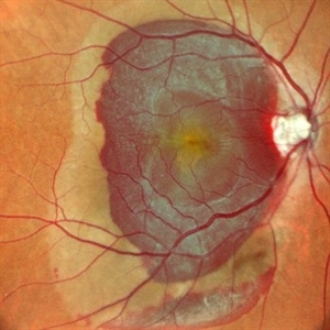
 Choroidal Neovascular Membrane (CNVM)
Sep 12 2023 by Ben Serar
Fundus photograph of LE showing Scarred CNVM at the macula, with sub-retinal fibrois with surrounding subretinal bleed
Condition/keywords: choroidal neovascular membrane (CNVM), scarring, sub-retinal fibrosis
-
 Choroidal Neovascularization
Mar 14 2015 by shah nasir
Images obtained from a 24-year-old man with blurred vision in the left eye with sub-retinal fluid (SRF) associated with choroidal neovessels (CNV). His visual acuity (VA) was 0.8 log MAR, sub-retinal fluid (SRF) was observed. Initial disrupted photoreceptor inner segment and outer segment junction (IS/OS) length was 2532µm, and choroidal neovascularization (CNV)thickness and diameter were 584 330µm respectively.
Photographer: Syed Nasir Ali Shah, Xian Jaiotong Medical University, Xian-Shaanxi 710061, China
Imaging device: CIRRUS HD-OCT
Condition/keywords: choroidal neovascularization (CNV)
-
 Choroidal Rupture with Choroidal Neovascularization
Jun 14 2021 by Shalin Shah
15-year-old boy with history of right eye blunt trauma presented with right eye choroidal rupture with choroidal neovascularization membrane(CNVM). OCT suggestive of CNVM with sub-retinal fluid.
Photographer: Shalin Shah, Dr Shroff's charity eye hospital, New Delhi, India
Imaging device: ZEISS VISUCAM 500
Condition/keywords: blunt trauma, choroidal neovascular membrane (CNVM), choroidal rupture
-
 Choroidal-rupture
Jan 2 2024 by Tahsin Khundkar, MD
37-year-old male with blunt ocular trauma presented with a choroidal rupture, pre -retinal and sub-retinal heme, and a heart shaped patch of commotio retinae.
Photographer: Jeffrey Zeigler, Concord Eye Center
Imaging device: Topcon
Condition/keywords: Choroidal Rupture, commotio retinae, Trauma
-
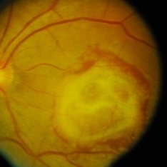
 Circumscribed Choroidal Hemangioma
Jul 3 2020 by Dhaivat Shah
A 30-year-old young male presented with drop in vision in right eye since 1 year (6/60). Fundus examination revealed choroidal hemangioma superotemporal to macula. Choroidal hemangioma is an unusual benign vascular tumor of the choroid. It can be circumscribed solitary or diffuse tumor with the later having other systemic associations. Circumscribed choroidal hemangiomas (CCHs) are usually unilateral, unifocal hamartomatous vascular tumor affecting people in second to fourth decade. It appers as round to oval, orangish-red mass in posterior pole with smooth homogenous surface mostly present in macular and peripapillary area. Hyperopic shift is seen in sub-foveal tumors in contrast to para-foveal ones which are usually asymptomatic or present with metamorphopsia or photopsia and diminished vision secondary to exudative retinal detachment. B-scan shows highly reflective tumor without any shadowing or acoustic solidity with high anterior A scan spike. EDI-OCT here depicts a smooth gently sloping choridal mass with compressed choriocapillaries and enlarged medium and large choroidal vessels. Over a period of time structural abnormalities of the outer retina can be visualised. Ancillary testing using Fluorescein Angiography shows lacy hyper-fluorescence during early arterial phase followed by increased hyper-fluorescence due to progressive profuse leakage from pin point foci during arterial and venous phase. Indocyanine green angiography shows lacy diffuse fluorescent tumor in early phase followed by hypo-fluorescent tumor due to dye wash out in late phase. Intrinsic auto-fluorescence is also seen in CCHs from lipofuscin and fresh sub-retinal fluid. Tumor is relatively hyper-intense with respect to vitreous in T1-weighted images in iso-intense in T2-weighted images of MRI. Asymptomatic cases need no treatment, while patients showing vision loss with presence or absence of exudative retinal detachment can be treated with photodynamic therapy which is preferred treatment due to site specific action. Selective occlusion of choroidal neovascularization can be achieved while the neurosensory retinal layers and Bruch membrane are almost unaffected, leaving retinal function intact. Green or rarely red wavelength laser photocoagulation is used to create a chorioretinal adhesion and resolve the SRF. Other treatment modalities include Transpupilary thermotherapy, external beam irradiation, proton beam therapy, brachytherapy and gamma knife.
Photographer: Miss Deepika Nagle
Imaging device: Zeiss
Condition/keywords: B scan ultrasound, choroidal hemangioma, fundus photograph, optical coherence tomography (OCT), photodynamic therapy
 Loading…