Search results (76 results)
-
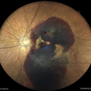
 Massive Sub-Retinal Haemorrhage involving Macula : Intra-operative still image
Oct 26 2023 by Veer Singh, MS, FVRS, FMRF, FICO (Retina)
Massive Sub-Retinal Haemorrhage involving Macula : Intra-operative still image
Photographer: Dr. Veer Singh
Imaging device: Ikegami 4k Microscope Camera
Condition/keywords: Haemorrhage, macula, Massive, Sub-Retinal
-
 Massive Subretinal Hemorrhage(
Apr 22 2020 by Mahesh Gopalakrishnan, MS, DO, DNB, FRCS
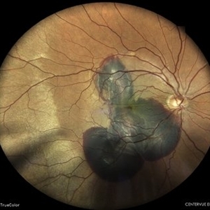
Optos ultra-widefield retinal image of the left eye of a 77-year-old male showing massive subretinal hemorrhage(SRH), subRPE hemorrhage and corrugated retinal appearance. He underwent tPA injection and vitrectomy with sub retinal blood drainage and silicone oil tamponade. Silicone oil removal was done after 7 months and final best corrected visual acuity was 20/50 .
Photographer: Rakesh PR , Giridhar Eye Institute, Kerala, India
Imaging device: Optos UWF Daytona Plus
Condition/keywords: sub-retinal pigment epithelium (RPE), subretinal hemorrhage
-
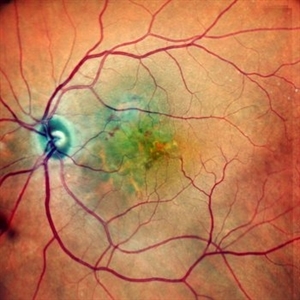
 Sub-Macular Haemorrhage
Apr 15 2023 by Veer Singh, MS, FVRS, FMRF, FICO (Retina)
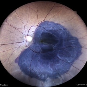
Sub-Retinal haemorrhage secondary to PCV in an elderly gentleman.
Photographer: Dr. Veer Singh
Condition/keywords: polypoidal choroidal vasculopathy (PCV), submacular hemorrhage
-
 Sub-Retinal Blood Air and TPA
Jan 31 2025 by Thirumalesh Mochi Basavaraj, MD
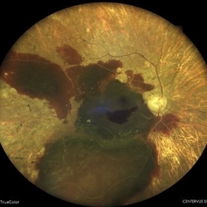
Intra Operative View of a 76 year old gentleman with Submacular bleed treated with Sub Retinal TPA, Ranibizumab and air, one can appreciate at multiple levels
Photographer: Thirumalesh Mochi Basavaraj
Condition/keywords: submacular hemorrhage, tissue plasminogen activator (tPA)
-
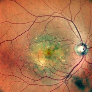
 SUB-RETINAL HEMORRHAGE
Feb 28 2023 by Akansha Sharma
COLOUR FUNDUS PHOTOGRAPH OF AN 84 YEAR OLD MALE WITH SUB RETINAL BLEED
Photographer: Dr. Urmil Shah, Dr. Denish Patel, Dr. Akansha Sharma, Bharati Eye Hospital, Ahmedabad, Gujarat
Condition/keywords: choroidal neovascular membrane (CNVM), subretinal hemorrhage
-
 SUB-RETINAL HEMORRHAGE WITH CHOROIDAL RUPTURE
Feb 28 2023 by Akansha Sharma
COLOUR FUNDUS PHOTOGRAPH OF A 45 YEAR OLD MALE WITH SUBRETINAL HEMORRHAGE WITH CHOROIDAL RUPTURE
Photographer: Dr. Urmil Shah, Dr. Denish Patel, Dr. Akansha Sharma, Bharati Eye Hospital, Ahmedabad, Gujarat
Condition/keywords: subretinal hemorrhage
-
 SUB-RETINAL HEMORRHAGE WITH FOVEOLAR DETACHMENT
Feb 28 2023 by Akansha Sharma
COLOUR FUNDUS PHOTO OF A 62 YEAR OLD FEMALE WITH SUB-RETINAL BLEED OPERATED WITH BIMANUAL VITRECTOMY WITH SUBRETINAL TPA INJECTION WITH SILICONE OIL INFUSION
Photographer: Dr. Akansha Sharma, Dr. Urmil Shah, Dr. Denish Patel, Eye Hospital, Ahmedabad, Gujarat
Condition/keywords: subretinal hemorrhage
-
 SUB-RETINAL HEMORRHAGE WITH RETINITIS PIGMENTOSA
Feb 28 2023 by Akansha Sharma
COLOUR FUNDUS PHOTOGRAPH OF A 77 YEAR OLD MALE WITH SUBRETINAL HEMORRHAGE IN A CASE OF RETINITIS PIGMENTOSA
Photographer: Dr. Urmil Shah, Dr. Denish Patel, Dr. Akansha Sharma, Bharati Eye Hospital, Ahmedabad, Gujarat
Condition/keywords: retinitis pigmentosa, subretinal hemorrhage
-
 SUB-RETINAL NEOVASCULAR MEMBRANE
Nov 21 2022 by Akansha Sharma
COLOUR FUNDUS PHOTO OF A 79 YEAR OLD MALE PATIENT WITH SUBRETINAL NEOVASCULAR MEMBRANE
Photographer: Dr. Akansha Sharma-Retina Foundation, Ahmedabad
Condition/keywords: choroidal neovascular membrane (CNVM), CNVM, subretinal neovascularization (SRNV)
-
 SUB-RETINAL NEOVASCULAR MEMBRANE
Nov 21 2022 by Akansha Sharma
COLOUR FUNDUS PHOTOGRAPH OF A 57 YEAR OLD MALE WITH SUBRETINAL NEOVASCULAR MEMBRANE
Photographer: Dr. Akansha Sharma-Retina Foundation, Ahmedabad
Condition/keywords: choroidal neovascularization (CNV), CNVM, subretinal neovascularization (SRNV)
-
 24 Hours Post Scleral Wound Closure+ Scleral Buckle+25 g Vitrectomy+Silicon Oil
Jan 23 2015 by Carlos Quezada-Ruiz, MD, FASRS
24 hours post op fundus photograph of a 43-year-old man who had perforating injury to the right eye with a small piece of plastic while he was hammering. OD LP, subconjunctival hemorrhage, clear cornea, hyphema, irido and ciclodyalisis as well as a luxated lens with traumatic cataract and a dense vitreous hemorrhage. B-US showed rhegmatogenous retinal detachment with a tear and a big inferior hemorrhagic choroidal detachment. 360 peritomy revealed 2-entry scleral wounds were found in zone II (M V and M VI) and closure was performed. 25 G PPV was performed with the infusion canal placed in the AC through the limbus. Lensectomy and removal of a dense recent vitreous hemorrhage revealed a white detached retina with an exit wound through the temporal inferior segment of the optic nerve with a nasal GRT and sub retinal hemorrhage as well as temporal inferior choroidal, PVD was induced and PFOs helped stabilizing the retina while vitrectomy and sub-retinal hemorrhage was removed through the GRT. Fluid air exchange was made and 360 endolaser over the buckle indentation was done and silicon oil was used as endotamponade. This picture was taken 24 hrs after the surgery.
Photographer: Lilibeth Rodriguez, Instituto de la Visión. Torreon, Mexico.
Condition/keywords: central retinal artery occlusion (CRAO), giant retinal tear, trauma
-
 Active CNVM
Jul 12 2023 by Harsh Vardhan Singh, MS
55-year male with left eye sub-retinal hemorrhage due to Active CNVM, Colour fundus photograph of left eye subretinal hemorrhage due to Active CNVM
Photographer: Harsh Vardhan Singh
Condition/keywords: choroidal neovascular membrane (CNVM), CNVM, subretinal hemorrhage
-
 Active CNVM
Jul 12 2023 by Harsh Vardhan Singh, MS
55-year male with left eye sub-retinal hemorrhage due to Active CNVM, Colour fundus photograph of left eye subretinal hemorrhage due to Active CNVM; Red-free image of left eye sub-retinal hemorrhage due to Active CNVM
Photographer: Harsh Vardhan Singh
Condition/keywords: choroidal neovascular membrane (CNVM), CNVM, subretinal hemorrhage
-
 Buckle Eroding Through to Subretinal Space
Apr 29 2015 by Philip J. Polkinghorne, MD
A high myope who underwent conventional retinal detachment surgery 20 years ago was noted to have an eroding scleral explant in the sub-retinal space.
Photographer: Janet Wigmore, Auckland Eye. Auckland, New Zealand.
Condition/keywords: extruding
-
 Central Serous Retinopathy
Mar 19 2024 by Corey Grant
Ultra Wide-Field Fundus Autofluorescence Imaging of a 37 year old female with Central Serous Retinopathy affecting her right eye. Patient Visual Acuity was 20/20 in both eyes. Patient reported black spots in her vision onset three years ago, with associating flashes of light. Patient reports history of cortisone back injections a few years ago and denies Flonase use. The physician stated that there is hyperautofluorescence in the area of gutter of Sub-Retinal Fluid which likely happened from CSR.
Photographer: Corey Grant, OSC
Imaging device: OPTOS CALIFORNIA RGB
Condition/keywords: Central Serous Chorioretinopathy (CSR), central serous retinopathy (CSR), fundus autofluorescence (FAF), Guttering, hyperautofluorescence, inferior retina, OPTOS, Retina, Right Eye, subretinal fluid, ULTRA WIDE FIELD
-
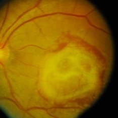
---thumb.JPG/image-square;max$300,300.ImageHandler) Choroidal Lymphoma
Choroidal Lymphoma
Nov 25 2012 by Mallika Goyal, MD
Left eye of a 60-year-old lady shows multiple sub-retinal yellowish masses. This is a recurrence 10 months following complete resolution of the lesions post-radiotherapy. Radiotherapy was repeated with regression of tumor.
Photographer: Mallika Goyal, MD, Apollo Health City, Hyderabad, India
-
 Choroidal Fracture
Oct 27 2024 by César Adrián Gómez Valdivia, MD
Fundus photograph of a traumatic choroidal fracture & extra-macular sub-retinal hemorrhage.
Photographer: @eyemissu2
Imaging device: TOPCON TRC-50DX
Condition/keywords: Choroidal Fracture
-
---thumb.JPG/image-square;max$300,300.ImageHandler) choroidal lymphoma
choroidal lymphoma
Nov 25 2012 by Mallika Goyal, MD
Left eye of a 60-year-old lady shows multiple sub-retinal yellowish masses of choroidal lymphoma. Radiotherapy resulted in complete regression with recurrence after 10 months.
Photographer: Mallika Goyal, MD, Apollo Health City, Hyderabad, India
Condition/keywords: lymphoma
-
 choroidal lymphoma
Nov 25 2012 by Mallika Goyal, MD
Left eye of a 60-year-old lady shows multiple sub-retinal yellowish masses of choroidal lymphoma. Radiotherapy resulted in complete regression with recurrence after 10 months.
Photographer: Mallika Goyal, MD, Apollo Health City, Hyderabad, India
Condition/keywords: lymphoma
-
---thumb.JPG/image-square;max$300,300.ImageHandler) Choroidal Lymphoma
Choroidal Lymphoma
Nov 25 2012 by Mallika Goyal, MD
Right eye of a 60-year-old lady shows multiple sub-retinal yellowish masses. This is a recurrence 10 months following complete resolution of the lesions post-radiotherapy. Radiotherapy was repeated with regression of tumor.
Photographer: Mallika Goyal, MD, Apollo Health City, Hydeabad, India
-
 Choroidal Neovascular Membrane (CNVM)
Sep 12 2023 by Ben Serar
Fundus photograph of LE showing Scarred CNVM at the macula, with sub-retinal fibrois with surrounding subretinal bleed
Condition/keywords: choroidal neovascular membrane (CNVM), scarring, sub-retinal fibrosis
-
 Choroidal Neovascularization
Mar 14 2015 by shah nasir
Images obtained from a 24-year-old man with blurred vision in the left eye with sub-retinal fluid (SRF) associated with choroidal neovessels (CNV). His visual acuity (VA) was 0.8 log MAR, sub-retinal fluid (SRF) was observed. Initial disrupted photoreceptor inner segment and outer segment junction (IS/OS) length was 2532µm, and choroidal neovascularization (CNV)thickness and diameter were 584 330µm respectively.
Photographer: Syed Nasir Ali Shah, Xian Jaiotong Medical University, Xian-Shaanxi 710061, China
Imaging device: CIRRUS HD-OCT
Condition/keywords: choroidal neovascularization (CNV)
 Loading…