Search results (76 results)
-
 Ocular Toxocariasis slide 1
Oct 22 2012 by Ronald C. Gentile, MD
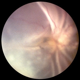
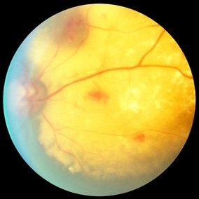
40-year-old man from South America was referred for a peripheral retinal scar in his left eye. He had a history of conjunctivitis as a child with exposure to multiple pets (cats and dogs). Fundus photo revealed a peripheral scarred sub-retinal granuloma located superior nasal with a retinal fold and traction extending to the optic nerve.
Photographer: The New York Eye & Ear Infirmary Department of Medical Imaging
Condition/keywords: toxocariasis
-
 Outer-Retinal-Tubulation
Jun 27 2013 by Jason S. Calhoun
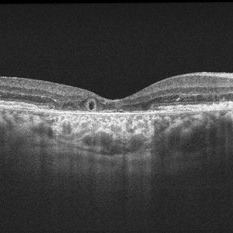
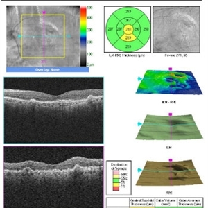
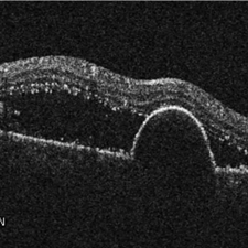
Patient with a history of wet macular degeneration and glaucoma in both eyes. VA is 20/50, right eye, 20/80, left eye. Patient is treated with Eylea in both eyes. Enhanced depth imaging OCT reveals a small like form of a cyst which in fact isn't a cyst at all. This is called outer retinal tubulation in which degenerating photo-receptors may become arranged in a circular or ovoid fashion. This is sometimes misdiagnosed as cystic changes in the retinal pigment epithelium or sub-retinal fluid.
Photographer: Jason S. Calhoun, Mayo Clinic Jacksonville, Florida
Imaging device: ZEISS OCT CIRRUS
Condition/keywords: optical coherence tomography (OCT)
-
 Ocular Toxocariasis slide 3
Oct 22 2012 by Ronald C. Gentile, MD
The sub-retinal scarred granuloma was white in color and elevated. It had pigment speckling around it. Serum testing was positive for past exposure to Toxocara canis.
Photographer: The New York Eye & Ear Infirmary Department of Medical Imaging
Condition/keywords: toxocariasis
-
 PVR Retinal Detachment with subretinal bands Slide 1
Oct 22 2012 by Ronald C. Gentile, MD
Total retinal detachment with pre-retinal and sub-retinal proliferation. The subretinal bands have a napkin ring configuration posteriorly with the macula folded and dragged above the optic nerve.
Photographer: The New York Eye & Ear Infirmary Department of Medical Imaging
Condition/keywords: proliferative vitreoretinopathy (PVR), subretinal bands
-
 Sub-RPE Hemorrhage, Trauma
Oct 18 2012 by Larry Halperin, MD
Sub-RPE hemorrhage from trauma
Condition/keywords: retinal pigment epithelium, sub-retinal pigment epithelium (RPE)
-
---thumb.JPG/image-square;max$300,300.ImageHandler) choroidal lymphoma
choroidal lymphoma
Nov 25 2012 by Mallika Goyal, MD
Left eye of a 60-year-old lady shows multiple sub-retinal yellowish masses of choroidal lymphoma. Radiotherapy resulted in complete regression with recurrence after 10 months.
Photographer: Mallika Goyal, MD, Apollo Health City, Hyderabad, India
Condition/keywords: lymphoma
-
 choroidal lymphoma
Nov 25 2012 by Mallika Goyal, MD
Left eye of a 60-year-old lady shows multiple sub-retinal yellowish masses of choroidal lymphoma. Radiotherapy resulted in complete regression with recurrence after 10 months.
Photographer: Mallika Goyal, MD, Apollo Health City, Hyderabad, India
Condition/keywords: lymphoma
-
 24 Hours Post Scleral Wound Closure+ Scleral Buckle+25 g Vitrectomy+Silicon Oil
Jan 23 2015 by Carlos Quezada-Ruiz, MD, FASRS
24 hours post op fundus photograph of a 43-year-old man who had perforating injury to the right eye with a small piece of plastic while he was hammering. OD LP, subconjunctival hemorrhage, clear cornea, hyphema, irido and ciclodyalisis as well as a luxated lens with traumatic cataract and a dense vitreous hemorrhage. B-US showed rhegmatogenous retinal detachment with a tear and a big inferior hemorrhagic choroidal detachment. 360 peritomy revealed 2-entry scleral wounds were found in zone II (M V and M VI) and closure was performed. 25 G PPV was performed with the infusion canal placed in the AC through the limbus. Lensectomy and removal of a dense recent vitreous hemorrhage revealed a white detached retina with an exit wound through the temporal inferior segment of the optic nerve with a nasal GRT and sub retinal hemorrhage as well as temporal inferior choroidal, PVD was induced and PFOs helped stabilizing the retina while vitrectomy and sub-retinal hemorrhage was removed through the GRT. Fluid air exchange was made and 360 endolaser over the buckle indentation was done and silicon oil was used as endotamponade. This picture was taken 24 hrs after the surgery.
Photographer: Lilibeth Rodriguez, Instituto de la Visión. Torreon, Mexico.
Condition/keywords: central retinal artery occlusion (CRAO), giant retinal tear, trauma
-
 Toxoplasmosis Slide 1
Oct 22 2012 by Ronald C. Gentile, MD
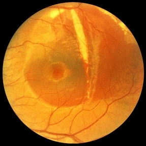
35-year-old women presented with decreasing vision in the left eye with progressive central scotoma. Fundus examination revealed one focal area of chorioretinitis adjacent to one of multiple old pigmented retinal scars. The focal area of chorioretinitis involved the deep retinal layers and was associated with sub-retinal fluid and little overlying vitritis.
Photographer: The New York Eye & Ear Infirmary Department of Medical Imaging
Condition/keywords: punctate outer retinal toxoplasmosis, toxoplasmosis
-
---thumb.JPG/image-square;max$300,300.ImageHandler) Choroidal Lymphoma
Choroidal Lymphoma
Nov 25 2012 by Mallika Goyal, MD
Right eye of a 60-year-old lady shows multiple sub-retinal yellowish masses. This is a recurrence 10 months following complete resolution of the lesions post-radiotherapy. Radiotherapy was repeated with regression of tumor.
Photographer: Mallika Goyal, MD, Apollo Health City, Hydeabad, India
Condition/keywords: lymphoma
-
---thumb.JPG/image-square;max$300,300.ImageHandler) choroidal lymphoma
choroidal lymphoma
Nov 25 2012 by Mallika Goyal, MD
Left eye of a 60-year-old lady shows multiple sub-retinal yellowish masses of choroidal lymphoma. Radiotherapy resulted in complete regression with recurrence after 10 months.
Photographer: Mallika Goyal, MD, Apollo Health City, Hyderabad, India
Condition/keywords: lymphoma
-
 Retinal Arterial Macroaneurysm
Sep 18 2018 by Somnath Chakraborty, MD
Left eye fundus photo of a 72-year-old hypertensive, female with a hemorrhagic retinal arterial macroaneurysm with sub-retinal blood.
Photographer: Pulok Chandra Roy, Retina Institute of Bengal
Condition/keywords: retinal arterial macroaneurysm
-
 Coats Disease Slide 1
Oct 22 2012 by Ronald C. Gentile, MD
A unilateral, sub-retinal, and yellowish exudative lesion with associated retinal telangiectasias involving the nasal retina. Refractile elements can be seen and represent cholesterol crystals.
Photographer: The New York Eye & Ear Infirmary Department of Medical Imaging
Condition/keywords: congenital retinal telangiectasis
-
 Multiple Retinal Lesions Secondary to Blunt Trauma
Jun 19 2018 by Somnath Chakraborty, MD
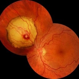
A montage of the right eye of a 15-year-old boy, who was struck by a football. The image shows multiple choroidal ruptures in the macular area, with sub-retinal blood and multiple, large retinal tears temporally. There is also an area of juxtapapillary, pigmentary changes.
Photographer: Saptarshi Mehta, Retina Institute of Bengal
Condition/keywords: blunt trauma, choroidal rupture, giant retinal tear, subretinal hemorrhage
-
---thumb.jpg/image-square;max$300,300.ImageHandler) Roth Spots and Retinal Hemorrhage
Roth Spots and Retinal Hemorrhage
Dec 27 2013 by David Callanan, MD
24-year-old patient, AML/ post-chemo thrombocytopenia with pre, intra, & sub-retinal.
Condition/keywords: white centered retinal hemorrhage (Roth Spot)
-
 Pars Plana Vitrectomy TPA 2
Oct 29 2012 by John S. King, MD
About 2 months after PPV, sub-retinal tPA, for the second time a large sub-retinal hemorrhage appeared. Visual acuity 20/40-20/50 after second ppv c subretinal tpa (vs cf prior to PPV), but less than result following first surgery (20/30).
Photographer: Kristin Konecki, OcuSight Eye Care Center, Rochester, NY
Imaging device: Cirrus
Condition/keywords: subretinal hemorrhage
-
 PVR, Vitreous Hemorrhage, Sub-retinal Debris
Dec 10 2012 by Yale L. Fisher, MD
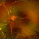
In this movie formed vitreous hemorrhage (yellow arrow) can be seen moving slowly inside this open-coned retinal detachment, which demonstrates no motion. ; Sub-retinal material (green arrow) with moderate reflectivity is seen moving in an undulating fashion slower than movement usually seen in the vitreous cavity.
Condition/keywords: video
-
 Retinal Pigment Epithelium Detachment
Jan 26 2016 by Andrea Arriola-Lopez, MD MSc
89-year-old woman, VA 20/800, IOP 13 mmHg. OCT showed subretinal fluid and PED. Anti-VEGF was administrated.
Photographer: Andrea Elizabeth Arriola MD, MSc
Imaging device: Cirrus
Condition/keywords: neovascular age-related macular degeneration (AMD), neovascularization (NV), sub-retinal pigment epithelium (RPE)
-
---thumb.JPG/image-square;max$300,300.ImageHandler) Choroidal Lymphoma
Choroidal Lymphoma
Nov 25 2012 by Mallika Goyal, MD
Left eye of a 60-year-old lady shows multiple sub-retinal yellowish masses. This is a recurrence 10 months following complete resolution of the lesions post-radiotherapy. Radiotherapy was repeated with regression of tumor.
Photographer: Mallika Goyal, MD, Apollo Health City, Hyderabad, India
-
---thumb.jpg/image-square;max$300,300.ImageHandler) Roth Spots and Retinal Hemorrhage
Roth Spots and Retinal Hemorrhage
Dec 27 2013 by David Callanan, MD
24-year-old patient, AML/ post-chemo thrombocytopenia with pre, intra, & sub-retinal.
Condition/keywords: white centered retinal hemorrhage (Roth Spot)
-
 Buckle Eroding Through to Subretinal Space
Apr 29 2015 by Philip J. Polkinghorne, MD
A high myope who underwent conventional retinal detachment surgery 20 years ago was noted to have an eroding scleral explant in the sub-retinal space.
Photographer: Janet Wigmore, Auckland Eye. Auckland, New Zealand.
Condition/keywords: extruding
-
 Tractional vs Combined Tractional/Rhegmatogenous Retinal Detachment with Active Neovascularization OS
Jun 1 2018 by Hosam Attia, MD
47-year-old African American, with history of diabetes mellitus of unknown duration and control, was referred for initial evaluation for conjunctival laceration in his left eye, following accidental finger nail injury, 6 days prior to presentation. - On exam, his vision was 20/50 OD and Bare HM/ LP OS. - Fundus color photos OD: No significant pathology, aside from attenuated vasculature OS: Chronic, Mac-Off, almost closed funnel tractional vs combined tractional/rhegmatogenous retinal detachment with large neovascularization (NVE) superiorly, detached ghost vessels, mild fresh vitreous hemorrhage, sub-retinal bands and inferior white vitreous debris from old hemorrhage (Not shown) - FA OD: No significant pathology aside from possible mild capillary non-perfusion in the extreme periphery, attenuated vasculature and possible tiny microaneurysms, nasally. OS: Extensive, wide spread capillary non- perfusion (correlate w/ detached Ghost vessels on color photos), and leakage from the NVE. - B/L Carotid Duplex was recommended due to the striking asymmetry in pathology with unknown medical history, diabetes duration and control etc (even in absence of any signs suggestive of possible ocular ischaemic syndrome OD)
Imaging device: Optos California
Condition/keywords: combined retinal detachment, tractional retinal detachment
-
 Buckle Eroding Through to Subretinal Space
Apr 29 2015 by Philip J. Polkinghorne, MD
A high myope who underwent conventional retinal detachment surgery 20 years ago was noted to have an eroding scleral explant in the sub-retinal space.
Photographer: Janet Wigmore, Auckland Eye. Auckland, New Zealand.
Condition/keywords: extruding
 Loading…