Search results (76 results)
-
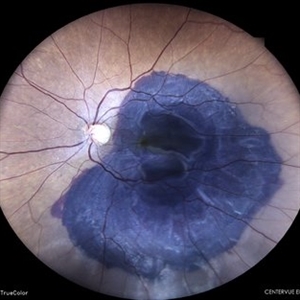
 Eye Finally Got the Ring... But the Retina Was Too Detached to Care
Nov 5 2025 by SHRADDHA RAJ SHRIVASTAVA
Left Eye B-scan ultrasound of a patient with old retinal detachment shows open funnel shaped hyperechoic membranous echoes, with high amplitude spikes on A-scan and a poor after-movement on dynamic B-scan, suggestive of retinal detachment. We can see a round echogenicity in sub-retinal location, with clear contents within, suggestive of a retinal cyst. This B-scan image is indicative of a long-standing chronic retinal detachment with secondary retinal cyst.
Photographer: Dr. Shraddha Raj Shrivastava
Condition/keywords: B scan ultrasound, chronic retinal detachment, OLD RD, open funnel RD, retinal cyst
-
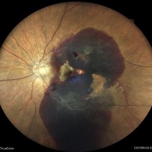
 Large Sub-retinal Band
Aug 15 2025 by Caleb Westhouse
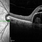
OCT and Optos imaging of a 58-year-old male with a macula-off retinal detachment, demonstrating a large subretinal band extending across the posterior pole. The OCT single-line scan provides a cross-sectional view of the subretinal band.
Photographer: Caleb Westhouse
Imaging device: Heidelberg Spectralis
Condition/keywords: Heidelburg Spectralis, OCT, Optos, Retinal detatatchment, Sub-retinal band
-
 Multi-modal Imaging of Type - 1 CNVM
May 30 2025 by Shivankar Sen, MS, FVRS
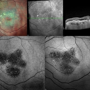
Multimodal Imaging of a case of Polypoidal Choroidal Vasculopathy Multicolor Reflectance showing multiple green-hyper-fringent lesions in the macular region (Up Left) Infra-red Autofluorescence and Blue Autofluorescence showing hypo-autofluorescent areas correspondingly revealing the exact extent of the sub-RPE Lesion (Down left and right respectively) Optical Coherence Tomography - Enhanced Depth Imaging showing Thumb-shaped Pigment Epithelial Detachment with presence of Sub-retinal fluid and Hyper-reflective foci (Top Right)
Photographer: Dr. Shivankar Sen
Imaging device: Heidelberg Spectralis HRA+OCT
Condition/keywords: Blue autofluroscence, CNVM, multicolor, near infrared autofluorescence (NIRAF), PCV, reflectance
-
 Diabetic Vitreous Hemorrhage
Feb 3 2025 by Hollie Sanders
61 year-old male with a history of type two DM. Per MD, Sub-Retinal, Pre-Retinal, and Vitreous Hemorrhages. Denies any treatment in the past. Treatment initiated in clinic.
Photographer: Hollie Sanders, Tennessee Retina, Nashville, Tennessee
Imaging device: OPTOS Silverstone Fundus camera
Condition/keywords: diabetic vitreous hemorrhage
-
 Sub-Retinal Blood Air and TPA
Jan 31 2025 by Thirumalesh Mochi Basavaraj, MD
Intra Operative View of a 76 year old gentleman with Submacular bleed treated with Sub Retinal TPA, Ranibizumab and air, one can appreciate at multiple levels
Photographer: Thirumalesh Mochi Basavaraj
Condition/keywords: submacular hemorrhage, tissue plasminogen activator (tPA)
-
 Galaxy
Oct 29 2024 by SHILPI H NARNAWARE, ICO ( Retina) , FAICO ( Vitreo-Retina)
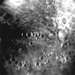
Retro mode image of a patient who underwent vitrectomy for RRD , with sub-retinal multiple PFCL bubbles giving appearance of arrangement of planets & other bodies in a galaxy
Photographer: Shilpi Narnaware, Sarakshi Netralaya , Nagpur, Maharashtra , India
Imaging device: Mirante ( by Nidek)
Condition/keywords: PFCL
-
 Choroidal Fracture
Oct 27 2024 by César Adrián Gómez Valdivia, MD
Fundus photograph of a traumatic choroidal fracture & extra-macular sub-retinal hemorrhage.
Photographer: @eyemissu2
Imaging device: TOPCON TRC-50DX
Condition/keywords: Choroidal Fracture
-
 Central Serous Retinopathy
Mar 19 2024 by Corey Grant
Ultra Wide-Field Fundus Autofluorescence Imaging of a 37 year old female with Central Serous Retinopathy affecting her right eye. Patient Visual Acuity was 20/20 in both eyes. Patient reported black spots in her vision onset three years ago, with associating flashes of light. Patient reports history of cortisone back injections a few years ago and denies Flonase use. The physician stated that there is hyperautofluorescence in the area of gutter of Sub-Retinal Fluid which likely happened from CSR.
Photographer: Corey Grant, OSC
Imaging device: OPTOS CALIFORNIA RGB
Condition/keywords: Central Serous Chorioretinopathy (CSR), central serous retinopathy (CSR), fundus autofluorescence (FAF), Guttering, hyperautofluorescence, inferior retina, OPTOS, Retina, Right Eye, subretinal fluid, ULTRA WIDE FIELD
-
 CSCR with Sub-Retinal Fibrin
Feb 6 2024 by Thirumalesh Mochi Basavaraj, MD
43 year old gentleman with a chronic central serous chorioretinopathy with sub retinal fibrin deposition.
Photographer: Puttaswamy
Condition/keywords: central serous chorioretinopathy (CSCR), subretinal fibrin deposition
-
 Choroidal-rupture
Jan 2 2024 by Tahsin Khundkar, MD
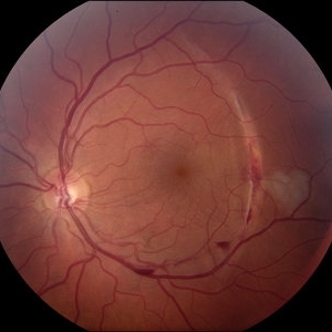
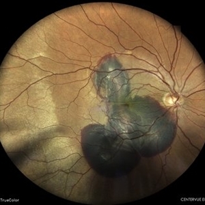
37-year-old male with blunt ocular trauma presented with a choroidal rupture, pre -retinal and sub-retinal heme, and a heart shaped patch of commotio retinae.
Photographer: Jeffrey Zeigler, Concord Eye Center
Imaging device: Topcon
Condition/keywords: Choroidal Rupture, commotio retinae, Trauma
-
 Layer Cake; Sub-retinal, Pre-retinal, Vitreous Hemorrhages
Dec 5 2023 by Virginia Gebhart
73 year old female with sub-retinal, pre-retinal, and vitreous hemorrhages all in OD. Will consider sx if blood does not clear on its own. Vision 20/40
Photographer: Virginia Gebhart
Imaging device: Topcon
Condition/keywords: pre-retinal hemorrhage, retinal macroaneurysm, subretinal hemorrhage, subretinal blood, vitreous hemorrhage
-
 Retinal detachment
Nov 23 2023 by Anand Temkar
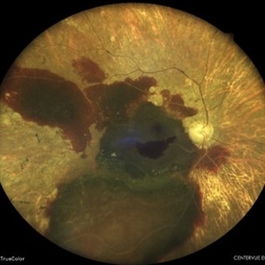
LE color photo montage of a 50 years old male with supero-nasal retinal detachment (with break) and we can see horseshoe tear temporally with sub-retinal fluid.
Photographer: Dr.Anand Temkar- Retina Foundation, Ahmedabad
Imaging device: Mirante
Condition/keywords: RD, retinal break
-
 Coat's disease
Nov 7 2023 by Harsh Vardhan Singh, MS
8-year-male with sub-retinal fibrosis with extensive exudates diagnosed with Coats' disease
Photographer: Harsh Vardhan Singh
Condition/keywords: Coats' disease
-
 Massive Sub-Retinal Haemorrhage involving Macula : Intra-operative still image
Oct 26 2023 by Veer Singh, MS, FVRS, FMRF, FICO (Retina)
Massive Sub-Retinal Haemorrhage involving Macula : Intra-operative still image
Photographer: Dr. Veer Singh
Imaging device: Ikegami 4k Microscope Camera
Condition/keywords: Haemorrhage, macula, Massive, Sub-Retinal
-
 Choroidal Neovascular Membrane (CNVM)
Sep 12 2023 by Ben Serar
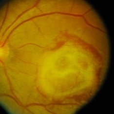
Fundus photograph of LE showing Scarred CNVM at the macula, with sub-retinal fibrois with surrounding subretinal bleed
Condition/keywords: choroidal neovascular membrane (CNVM), scarring, sub-retinal fibrosis
-
 Neovascular AMD with Ring Shaped lesions
Jul 12 2023 by Gregg T. Kokame, MD, MMM, FASRS
Horizontal OCT Scan - Neovascular AMD with Active CNV Ring shaped lesions underneath the RPE inverted U-shaped elevation
Photographer: Jaclyn Pisano
Imaging device: Zeiss Cirrus 6000
Condition/keywords: inverted u-shaped elevation, macular edema, OCT, Sub-retinal fluid, wet age-related macular degeneration (wet AMD)
-
 Neovascular AMD with Ring Shaped lesions
Jul 12 2023 by Gregg T. Kokame, MD, MMM, FASRS
Vertical OCT Scan - Neovascular AMD with Active CNV Ring shaped lesions underneath the RPE inverted U-shaped elevation
Photographer: Jaclyn Pisano
Imaging device: Zeiss Cirrus 6000
Condition/keywords: edema, OCT, Sub-retinal fluid, wet age-related macular degeneration (wet AMD)
-
 Active CNVM
Jul 12 2023 by Harsh Vardhan Singh, MS
55-year male with left eye sub-retinal hemorrhage due to Active CNVM, Colour fundus photograph of left eye subretinal hemorrhage due to Active CNVM; Red-free image of left eye sub-retinal hemorrhage due to Active CNVM
Photographer: Harsh Vardhan Singh
Condition/keywords: choroidal neovascular membrane (CNVM), CNVM, subretinal hemorrhage
-
 Active CNVM
Jul 12 2023 by Harsh Vardhan Singh, MS
55-year male with left eye sub-retinal hemorrhage due to Active CNVM, Colour fundus photograph of left eye subretinal hemorrhage due to Active CNVM
Photographer: Harsh Vardhan Singh
Condition/keywords: choroidal neovascular membrane (CNVM), CNVM, subretinal hemorrhage
-
 Sub-Macular Haemorrhage
Apr 15 2023 by Veer Singh, MS, FVRS, FMRF, FICO (Retina)
Sub-Retinal haemorrhage secondary to PCV in an elderly gentleman.
Photographer: Dr. Veer Singh
Condition/keywords: polypoidal choroidal vasculopathy (PCV), submacular hemorrhage
-
 Pigment Epithelium Detachment, Secondary to AMD
Mar 17 2023 by Ceara Donovan
Optical coherence tomography of a 76 year old woman with a Pigment Epithelium Detachment, Secondary to AMD affecting her right eye. Patient had no significant response to Avastin, Eylea, Lucentis 0.5, or Vabysmo and was switched to Beovu. Following Beovu intravitreal injection her edema improved on OCT. Patient's vision was sc20/200+1 at the time the image was taken.
Photographer: Ceara Donovan
Imaging device: Heidelberg Spectralis
Condition/keywords: exudative age-related macular degeneration, heidelberg spectralis, macular degeneration, optical coherence tomography (OCT), pigment epithelial detachment (PED), Sub-retinal fluid
-
 SUB-RETINAL HEMORRHAGE WITH RETINITIS PIGMENTOSA
Feb 28 2023 by Akansha Sharma
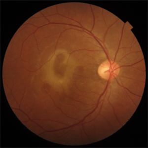
COLOUR FUNDUS PHOTOGRAPH OF A 77 YEAR OLD MALE WITH SUBRETINAL HEMORRHAGE IN A CASE OF RETINITIS PIGMENTOSA
Photographer: Dr. Urmil Shah, Dr. Denish Patel, Dr. Akansha Sharma, Bharati Eye Hospital, Ahmedabad, Gujarat
Condition/keywords: retinitis pigmentosa, subretinal hemorrhage
-
 SUB-RETINAL HEMORRHAGE WITH CHOROIDAL RUPTURE
Feb 28 2023 by Akansha Sharma
COLOUR FUNDUS PHOTOGRAPH OF A 45 YEAR OLD MALE WITH SUBRETINAL HEMORRHAGE WITH CHOROIDAL RUPTURE
Photographer: Dr. Urmil Shah, Dr. Denish Patel, Dr. Akansha Sharma, Bharati Eye Hospital, Ahmedabad, Gujarat
Condition/keywords: subretinal hemorrhage
-
 SUB-RETINAL HEMORRHAGE WITH FOVEOLAR DETACHMENT
Feb 28 2023 by Akansha Sharma
COLOUR FUNDUS PHOTO OF A 62 YEAR OLD FEMALE WITH SUB-RETINAL BLEED OPERATED WITH BIMANUAL VITRECTOMY WITH SUBRETINAL TPA INJECTION WITH SILICONE OIL INFUSION
Photographer: Dr. Akansha Sharma, Dr. Urmil Shah, Dr. Denish Patel, Eye Hospital, Ahmedabad, Gujarat
Condition/keywords: subretinal hemorrhage
-
 SUB-RETINAL HEMORRHAGE
Feb 28 2023 by Akansha Sharma
COLOUR FUNDUS PHOTOGRAPH OF AN 84 YEAR OLD MALE WITH SUB RETINAL BLEED
Photographer: Dr. Urmil Shah, Dr. Denish Patel, Dr. Akansha Sharma, Bharati Eye Hospital, Ahmedabad, Gujarat
Condition/keywords: choroidal neovascular membrane (CNVM), subretinal hemorrhage
 Loading…