Search results (189 results)
-
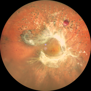
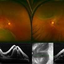
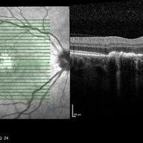
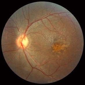
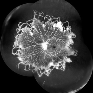 Vascular Non Perfusion in Takayasu Arteritis
Feb 6 2024 by SHILPI H NARNAWARE, ICO ( Retina) , FAICO ( Vitreo-Retina)
A case of 16 year-old female with combined RD in RE. Fundus examination & FFA revealed 360 degrees non-perfusion in periphery in non-symptomatic eye.
Photographer: Shilpi Narnaware, Sarakshi Netralaya , Nagpur, Maharashtra , India
Imaging device: Mirante ( by Nidek)
Condition/keywords: CNP areas, takayasu arteritis
-
 Vitreous Cavity Inhabitant
Jun 2 2025 by Poornachandra B, MS, FVRS
A 36-year-old male presented with a 6-week history of intermittent ocular redness, now accompanied by the recent onset of floaters for the past 2 days. Fundus examination revealed the presence of a nematode in the vitreous cavity.
Photographer: Mr Dhikshith
Condition/keywords: parasite
-
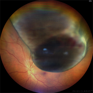
 Hourglass in an Eye
Apr 22 2025 by KRISHNENDU NANDI, MS
A twenty-five-year-young male presented with a decrease in vision in the right eye following a blunt trauma with a football. On examination the BCVA in the right eye was CFCF and the left eye was 6/6, N6. The anterior segment was within normal limits. AT was 12 and 10 mm of Hg in the right and left eyes, respectively. Fundus examination reveals subhyaloid haemorrhage in the right eye with an attached retina. The fundus of the left eye was within normal limits. YAG laser hyaloidotomy was done with an energy of 2 mJ in the right eye. After 3 weeks the BCVA in the right eye improved to 6/9, N6.
Photographer: Dr. Krishnendu Nandi
Imaging device: Topcon
Condition/keywords: Trauma, YAG HYALOIDOTOMY, Young Male
-
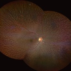
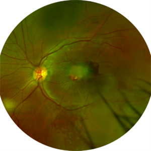
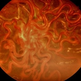 Wyburn-Mason Syndrome (Racemose Angioma)
Mar 23 2024 by Pushkar Mahale
Fundus photograph of a 10 year old child presenting with no perception of light in right eye. Fundus examination revealed dilated and tortuous retinal vessels suggestive of Racemose Hemangioma.
Photographer: Dr Pushkar Mahale
Condition/keywords: racemose hemangioma, Wyburn -Mason Syndrome
-
 Acute Syphilitic Posterior Placoid Chorioretinitis
May 4 2021 by RAFAEL REIS PEREIRA, MD
A 31-year-old patient with a complaint of photophobia and low visual acuity OD in the previous three weeks. BCVA was 20/60 and 20/20 The fundus examination revealed a placoid white lesion in the posterior pole and vitreous cells in the right eye. The left eye was unremarkable. Fluorescein angiography reveals hyperfluorescent plaque with distinctive “leopard spots” hypofluorescence.
Imaging device: Opto California
Condition/keywords: acute syphilitic posterior placoid chorioretinitis
-
 Annular Tractional Retinal Detachment
Jul 4 2024 by Hector Gabriel Moreno Solano, MD, MHA
52-year-old Hispanic female patient with a diagnosis of type II diabetes mellitus of 15 years of evolution, comes to the retina service for progressive visual loss in the right eye (single functional eye) with visual acuity of 20/100, Fundus examination reveals laser-modified proliferative diabetic retinopathy with activity + annular tractional retinal detachment with macular involvement.
Photographer: Hector Gabriel Moreno Solano, MD, MHA, HGZ #20 IMSS Puebla.
Imaging device: Mirante
Condition/keywords: macular detachment, proliferative diabetic retinopathy (PDR), tractional retinal detachment
-
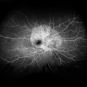
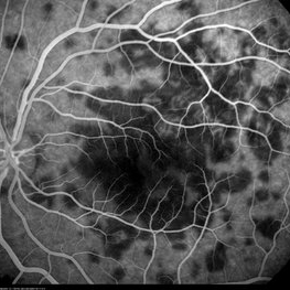 APMPPE in a 21-Year-Old Female Patient
Oct 23 2015 by Roy Schwartz, MD
FA photograph of a 21-year-old, usually healthy, female, presenting with visual deterioration and photophobia in BE. Upon examination deep lesions were seen on fundus examination. FA showed hypofluorescent lesions (seen here at 36 seconds) that later became hyperfluorescent
Photographer: Galit Yair Pur
Condition/keywords: acute posterior multifocal placoid pigment epitheliopathy (APMPPE)
-
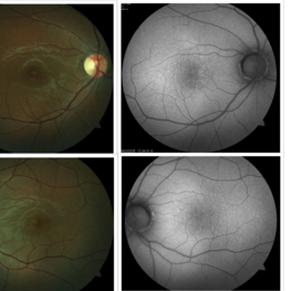 Bilateral Benign Yellow Dot Maculopathy
May 6 2025 by Amol yuvraj ganvir
A 37-year-old female patient presented for a routine eye examination. Her best-corrected visual acuity was 6/6 in both eyes. Fundus examination revealed multiple small yellow dots over the macula in both eyes. FAF imaging demonstrated characteristic hyperautofluorescence corresponding to these dots.
Photographer: Dr. Amol Ganvir, Vitreo-Retina Fellow, Ishwar Eye Centre, Rohtak, Haryana
Imaging device: Visucam-Zeiss
Condition/keywords: Autoflourescence, yellow dots
-
---thumb.jpg/image-square;max$300,300.ImageHandler) Birdshot Case #1 OD IVFA
Birdshot Case #1 OD IVFA
May 1 2013 by Armando L. Oliver, MD
64-year-old Puerto Rican woman consulted due to the presence of 1+ vitreous cells. The fundus examination revealed orange to yellow lesions dispersing from the disk. Work-up revealed she was HLA-A29 positive and the suspected diagnosis of Birdshot Chorioretinopathy was made. Chest X-Ray, FTA-Abs and RPR were negative.
Photographer: Moises Castro, Instituto de Ojos y Piel, Carolina, PR
Imaging device: Zeiss, Visucam NM/FA
Condition/keywords: birdshot, birdshot chorioretinopathy, birdshot retinochoroidopathy
-
 Chronical Submacular Hemorrhage in the Setting of Neovascular AMD
Mar 23 2015 by Rita Couceiro, MD, MS
An 80-year-old male, with a history of hypertension and high cholesterol, complained of acute and painless vision loss in his left eye (OS) in the previous 5 months. On observation best corrected visual acuity in OS was hand motion. A dense vitreous opacity in OS precluded fundus examination. Ocular ultrasound revealed vitreous hemorrhage and thickening of the macular area. The patient was submitted to pars plana vitrectomy, which disclosed a large submacular hemorrhage with chronical features and disciform scarring in the setting of neovascular AMD.
Imaging device: Intraoperative fundus photograph
Condition/keywords: neovascular age-related macular degeneration (AMD), submacular hemorrhage, wet age-related macular degeneration (wet AMD)
-
 Dislocated Lens
Jun 29 2013 by Jason S. Calhoun
84-year-old female comes in with blurred vision in the left eye. VA was 20/30, right eye and count fingers in the left eye. Fundus examination reveals dislocation of the IOL into the vitreous inferiorily at 6-o'clock. Suggest surgery to fix the problem.
Photographer: Jason S. Calhoun, Mayo Clinic Jacksonville, Florida
Imaging device: TOPCON TRC 50-EX
Condition/keywords: dislocated posterior chamber intraocular lens (PCIOL)
-
 ERM
ERM
Nov 26 2020 by Priya Rasipuram Chandrasekaran, MBBS, DO, DNB, FRCS
A 58-year-old female presented with distortion of images 1 month following cataract surgery in the right eye and fundus examination showed epiretinal membrane extending from the disc to the macula and OCT macula showing epiretinal membrane with disorganization of the foveal architecture.
Condition/keywords: epiretinal membrane (ERM)
-
 Fundus Albipunctatus
Aug 25 2022 by Aditya S Kelkar, MS, FRCS, FASRS,FRCOphth
65 year old female, presented for cataract evaluation. Fundus examination showed whitish-yellow flecks in the retina.
Imaging device: Clarus 500
Condition/keywords: fundus albipunctatus, fundus photograph
-
 Hypertensive Retinopathy Pre
Mar 10 2014 by Dong Yoon Kim, MD
20-year-old women who had severe preeclamsia visited our clinic for decreased visual acuity on her both eyes. Her visual acuity was 20/100 on both eyes. Fundus examination revealed serous retinal detachment on both eyes. OCT examination revealed subretinal and intraretinal fluid.
Photographer: Sun Tae Kim, University of Ulsan, Asan Medical Center
Imaging device: Optos C200 MA scanning laser ophthalmoscope
Condition/keywords: hypertensive retinopathy
-
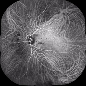 Multifocal Choroiditis
Aug 16 2018 by FELIPE PEREIRA
Mid-phase indocyanine green angiography of a 25-year-old woman with sudden central vision loss and photopsias for 7 days. The hypofluorescent lesions in the macula and nasal to the disc correspond to the yellow-white deep lesions in the fundus examination. No leakage is observed at any stage of the exam
Photographer: Claudio Zett Lobos
Imaging device: HEIDELBERG SPECTRALIS HRA
Condition/keywords: indocyanine green (ICG) angiography, multifocal choroiditis, white dot syndrome
-
 Peri-papillary Vascular Loop
Jun 2 2020 by Dhaivat Shah
Peri-papillary vascular loops (PVL) are rare congenital vascular malformations, which are usually detected as accidental finding during routine fundus examination. They can often be confused with tributary vein occlusion or racemose hemangioma. Although benign and asymptomatic, they can be rarely associated with vitreous hemorrhage and arterial occlusion. We herein present a case of a 60-year-old hypertensive male, who was diagnosed elsewhere to have a tributary vein occlusion and was referred to us. FFA was advised to rule out neovascularization, surrounding capillary non perfusion and mass lesion (hemangioma). On FFA, the arterial loop showed a slightly delayed filling (3-5 seconds) as compared to the other arterial vessels and the original vessel appeared to be a branch arising from central retinal artery. The choroidal filling was delayed in the area supplied by the loop. A cilioretinal artery was also noted. The patient was diagnosed to have a Peri-papillary vascular arterial loop (PVL), likely to be congenital in origin. The patient was reassured and was advised yearly follow up. These loops are usually accidental findings discovered during routine fundus examination. Since these vessels are looped and tortuous, they exhibit a slower and laminar blood flow, which make them more prone for arterial occlusions. The vitreous in this area tends to be adherently attached, so during PVD induction, it is likely to cause a tear and hemorrhage leading to vitreous hemorrhage. Until and unless there is a break, this hemorrhage tends to resolve on its own and does not warrant treatment. If there is an evident break, it can be dealt with laser barrage.
Photographer: Choithram Netralaya
Condition/keywords: congenital prepapillary vascular loop
-
 Serpiginous Choroidopathy
Sep 24 2024 by Gustavo Uriel Fonseca Aguirre
Right fundus of a 32-year-old female patient diagnosed with serpiginous choroiditis.
Photographer: Gustavo U. Fonseca Aguirre, Fundación Hospital Nuestra Señora de la Luz, Ciudad de México
Condition/keywords: Fundus examination, serpiginous choroiditis
-
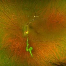
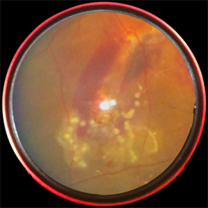 Subretinal Worm with Laser Marks - Smartphone Fundus Photograph
Jun 13 2019 by Prithvi Chandrakanth
42-year-old, male came with chief complaints of diminished vision and floaters in right eye for past one week. On fundus examination noted to have subretinal haemorrhage and edema at the posterior pole and a subretinal live mobile worm at the periphery. Laser photocoagulation done followed by pars plana vitrectomy.
Photographer: Dr.PRITHVI CHANDRAKANTH, Dr.CHANDRAKANTH MALABAR NETHRALAYA, KOZHIKODE
Imaging device: TRASH TO TREASURE RETCAM - SMARTPHONE FUNDUS CAMERA DEVICE
Condition/keywords: laser photocoagulation, smartphone fundus photography, subretinal hemorrhage, uveitis, worm
-
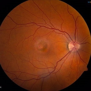
 Unilateral Acute Idiopathic Maculopathy OCT Macula
May 7 2019 by William Ensor
A 37-year-old female presented with a two-week history of vision loss in the right eye. She experienced a flu-like illness including rash on the hands, feet, and mouth 2 days prior to her vision change. Her 3-year-old son had a similar illness diagnosed as hand, foot, and mouth disease by his pediatrician one week prior. Her visual acuity was 20/150 of the right eye, and 20/20 of the left eye. On dilated fundus examination, the left eye was unremarkable; the right eye revealed a circular, variably pigmented lesion of the macula. OCT imaging showed areas of RPE loss and clumping, with overlying loss of the photoreceptor layer. Fluorescein angiography showed central and peripheral hyperfluorescence consistent with window defect, and blockage in area of RPE loss. No treatment was initiated at this time. The patient returned 10 days later; her visual acuity improved to 20/50 in the right eye. Dilated fundus exam showed increased pigmentation of the macular lesion. OCT of the right eye showed further RPE clumping without recovery of the photoreceptor layer, despite her improved visual acuity.
Condition/keywords: unilateral acute idiopathic maculopathy
-
 Uveal Melanoma
Apr 26 2025 by Vishal Agrawal, MD, FRCS,FACS,FASRS
A 32 year-old male presented with complaints of perceiving a shadow in OS for 15-20 days. His BCVA was 20/20 OU. On Fundus examination, a large, elevated, well-defined, pigmented choroidal mass with few hemorrhages over the lesion was seen and a provisional diagnosis of uveal melanoma was made. urgent oncological consultation was recommended for further treatment.
Photographer: Dr Ayushi Gupta
Imaging device: Clarus 700
Condition/keywords: melanoma
-
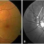
 Unilateral Acute Idiopathic Maculopathy Fundus
May 7 2019 by William Ensor
A 37-year-old female presented with a two-week history of vision loss in the right eye. She experienced a flu-like illness including rash on the hands, feet, and mouth 2 days prior to her vision change. Her 3-year-old son had a similar illness diagnosed as hand, foot, and mouth disease by his pediatrician one week prior. Her visual acuity was 20/150 of the right eye, and 20/20 of the left eye. On dilated fundus examination, the left eye was unremarkable; the right eye revealed a circular, variably pigmented lesion of the macula. OCT imaging showed areas of RPE loss and clumping, with overlying loss of the photoreceptor layer. Fluorescein angiography showed central and peripheral hyperfluorescence consistent with window defect, and blockage in area of RPE loss. No treatment was initiated at this time. The patient returned 10 days later; her visual acuity improved to 20/50 in the right eye. Dilated fundus exam showed increased pigmentation of the macular lesion. OCT of the right eye showed further RPE clumping without recovery of the photoreceptor layer, despite her improved visual acuity.
Condition/keywords: unilateral acute idiopathic maculopathy
-
 Combined Hamartoma
Feb 29 2016 by Andrea Arriola-Lopez, MD MSc
40 year-old man with diminished VA since 6 month ago. Fundus examination revealed macular folds, yellow-whitish elevated lesion at the fovea and a subretinal hemorrhage.
Photographer: Andrea Elizabeth Arriola-Lopez MD, MSc
Imaging device: OPTOS Dakota
Condition/keywords: combined hamartoma, macula, subretinal hemorrhage
-
 Cone Rod Dystrophy slide 2
Oct 22 2012 by Ronald C. Gentile, MD
Fundus examination revealed spotty pigmentary changes in the macular area with some peripheral depigmentation of the retinal pigment epithelium.
Photographer: The New York Eye & Ear Infirmary Department of Medical Imaging
Condition/keywords: cone dystrophy, retinal pigment epithelium
-
 Hemangioma
Oct 16 2012 by Anat Loewenstein, MD
Fundus examination of a 68 year old lady with decreased vision in her left eye for several months. VA 20/30 in her RE and counting fingers in the left eye. In the left eye there was a large red mass protruding and covering almost the entire optic nerve. Diagnosed as retinal hemangioma. The patient underwent low fluence PDT.
Photographer: Galit Yair-Pur
Condition/keywords: hemangioma
-
 Ocular Hypotony Due to Leaking Bleb
Apr 1 2019 by Anfisa Ayalon, MD
81-year-old male who had trabeculectomy in his right eye 4 years ago, presented to the emergency room with complains of decreased vision in that eye for two months. Slit-lamp examination showed cystic bleb with leakage, intraocular pressure was 0 MMHg. Fundus examination showed hypotony maculopathy, peripheral choroidal detachments, multiple chorioretinal folds with subretinal fluid.
Photographer: Anfisa Ayalon, MD., Meir Medical Center, Kfar Saba, Israel.
Imaging device: California, Optos 200 DTX
Condition/keywords: choroidal detachment, hypotonous retinopathy, hypotony maculopathy
 Loading…