File number: 28479
Comments
-
 Suber S. Huang, MD, MBA, FASRS (September 21 2018)
Superb capture! It would be good to know when this image was captured in relation to her symptoms. Interesting that there was no ICG leakage. Perhaps in the recovery phase? Consider adding to clinical history and add ICG as a key word. Thank you!
Sign in to comment.
 Initializing download.
-
 By FELIPE PEREIRA
UNIFESP
Co-author(s): Claudio Zeet Lobos - Uploaded on Aug 16, 2018.
- Last modified by FELIPE PEREIRA on Dec 22, 2018.
- Rating
- Appears in
- Multifocal Choroiditis
- Condition/keywords
- white dot syndrome, multifocal choroiditis, indocyanine green (ICG) angiography
- Photographer
- Claudio Zett Lobos
- Imaging device
- HEIDELBERG SPECTRALIS HRA
- Description
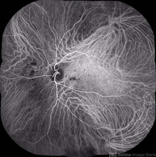
- Mid-phase indocyanine green angiography of a 25-year-old woman with sudden central vision loss and photopsias for 7 days. The hypofluorescent lesions in the macula and nasal to the disc correspond to the yellow-white deep lesions in the fundus examination. No leakage is observed at any stage of the exam
")


")
")
")
")

")