File number: 15179
Comments
-
 James B. Soque, CRA, OCT-C, COA, FOPS (March 11 2014)
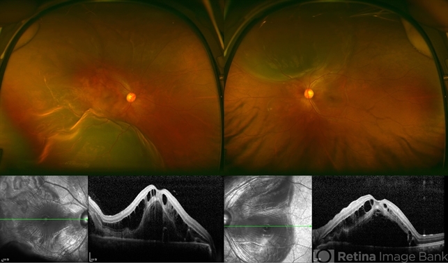
Dr Kim has created a beautiful montage of the Optos 200 SLO, and it is well documented in this case of hypertension secondary to her preeclamsia. What is important is that this kind of documentation of retinal detachment is most useful when there are details visible on the OLM, and other peripheral retinal layers at the detachment point(s), visible with the OCT. 200 degrees of field of view is very useful in planning surgery for her bilateral RD.
Sign in to comment.
 Initializing download.
-
 By Dong Yoon Kim, MD
Top Retina Center
Co-author(s): Young Hee Yoon, University of Ulsan, Asan Medical Center - Uploaded on Mar 10, 2014.
- Last modified by Caroline Bozell on Mar 10, 2014.
- Rating
- Appears in
- Fungal endoopthalmitis
- Condition/keywords
- hypertensive retinopathy
- Photographer
- Sun Tae Kim, University of Ulsan, Asan Medical Center
- Imaging device
-
Scanning laser ophthalmoscope
Optos C200 MA scanning laser ophthalmoscope - Description
- 20-year-old women who had severe preeclamsia visited our clinic for decreased visual acuity on her both eyes. Her visual acuity was 20/100 on both eyes. Fundus examination revealed serous retinal detachment on both eyes. OCT examination revealed subretinal and intraretinal fluid.


---thumb.JPG/image-square;max$79,0.ImageHandler "hypertensive retinopathy")
---thumb.JPG/image-square;max$79,0.ImageHandler "hypertensive retinopathy")


---thumb.JPG/image-square;max$79,0.ImageHandler "Hypertensive Retinopathy")
