 Initializing download.
-
 By Jessilla Phou
Retina and Uveitis Center
Co-author(s): Muge Kesen, MD - Uploaded on Jul 10, 2025.
- Last modified by Joshua Friedman on Jul 11, 2025.
- Rating
- Appears in
- 10-Jul-2025
- Condition/keywords
- fundus photograph, cherry red spot, Sialidosis
- Photographer
- Jessilla Phou
- Imaging device
-
Fundus camera
Optos California - Description
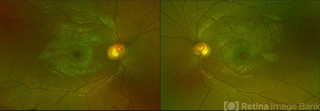
- These are fundus photographs capturing an 18 year old male with Type 1 Sialidosis, a rare inherited lysosomal storage disorder caused by a deficiency in the neuraminidase 1 (Neu1) enzyme. Currently, there are fewer than 1,000 people in the USA who have this disorder. It is characterized by a cherry red spot in the macula which occurs when lipids accumulate in the retinal ganglion cells. This causes the macula to appear red as seen in these fundus images. The patient presented at our office with ataxia, depth perception issues, and slow reaction time. His visual acuity was 20/40, suggestive of early stage Sialidosis.
")
")
")


