 Initializing download.
-
 By Anand Temkar
Co-author(s): Dr.Surendra Pal- Vasan Eye Hospital, Madurai - Uploaded on Sep 7, 2025.
- Last modified by Joshua Friedman on Sep 8, 2025.
- Rating
- Appears in
- RVO
- Condition/keywords
- venous occlusion, macular branch retinal vein occlusion (BRVO)
- Photographer
- Dr.Anand Temkar- Vasan Eye Hospital, Tiruchirappalli
- Imaging device
-
Optical coherence tomography system
Opticon - Description
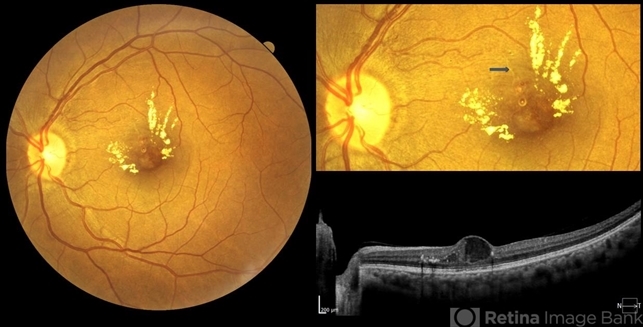
- A 55 yrs old female, k/c/o DM ( type II ) since past 5 yrs ( on medication ). Her vision was 6/6 in RE and 6/24 in her LE. IOP was 16 in both eyes. On examination, RE was WNL, and in LE ( color photo ) we noticed exudates, small hemorrhages, edema and sclerosed vessel ( depicted by black arrow. OCT LE shows altered foveal contour with cystic spaces and intraretinal hyperreflective material ( IRHRM ).


