 Initializing download.
-
 By Virginia Gebhart
Retina Consultants of Carolina
Co-author(s): Chris Bergstrom, MD, OD - Uploaded on Jul 16, 2025.
- Last modified by Virginia Gebhart on Aug 15, 2025.
- Rating
- Appears in
- Miscellaneous
- Condition/keywords
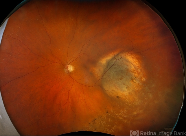
- choroidal melanoma, amelanotic melanoma
- Photographer
- Virginia Gebhart, Retina Consultants of Carolina
- Imaging device
-
Fundus camera
Optos California - Description
- 78 year old male with a partially amelanotic dome-shaped lesion with RPE changes, hard exudates, overlying intraretinal fluid and minimal SRF temporally. Exam and ultrasound findings consistent with choroidal melanoma. Pt will be scheduled for brachytherapy pending CT scan results.


