Search results (569 results)
-
 LCA type 12
Apr 10 2025 by Joshua Friedman
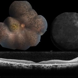
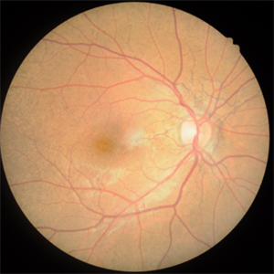
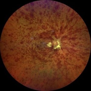
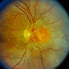
LCA type 12 due to pathogenic mutations in RDH12. 13-year-old male with a visual acuity of 20/80 and 20/300 in the right and left eye, respectively. There is extensive pigment migration in the peripheral retina and macula. Like RPE65, there is widespread hypoautofluorescent signal, however, the peripapillary retina is uniquely spared in this form of LCA. On OCT, there is almost complete loss of the retina centrally.
Photographer: Stephen Tsang, MD, PhD
Condition/keywords: Leber Congenital Amaurosis
-
 Multimodal Imaging in CHRPE
Mar 6 2025 by Gerardo - Montante Montelongo, MD
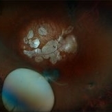
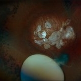
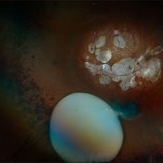
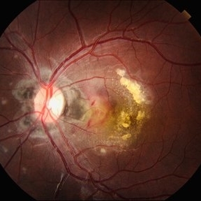
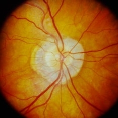
Fundus photograph of an 83-year-old male with a history of Diabetes, smoking, cataract surgery on the right eye in 2022, and open-angle glaucoma. Asymptomatic. Indirect ophthalmoscopy revealed 80% excavation, peripapillary atrophy, and a hyperpigmented perifoveal lesion with 35% atrophy, 10% drusen, and 5.1 mm diameter, corresponding to a CHRPE. At multimodal imaging, FFA shows hypoautofluorescence of the lesion, OCT shows preservation of internal retinal layers, atrophy of external retinal layer, with an RPE disruption, and posterior shadowing. USG shows a flat hyperechoic lesion 5.1 mm in diameter and 1.32 mm in thickness, solid and with high internal reflectance.
Photographer: Gerardo Montante-Montelongo, MD, Mexican Institute of Ophthalmology
Imaging device: Clarus 700
Condition/keywords: congenital hypertrophy of the retinal pigment epithelium (CHRPE), multimodal imaging
-
 Maternally-Inherited Diabetes and Deafness (MIDD) Syndrome
Jan 12 2025 by Niloofar Piri, MD
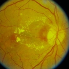
Fundus Autofluorescence image of right posterior pole in a 43 year old female who was referred for diabetic retinopathy evaluation, demonstrated multiple patches of hypoautrofluorescence surrounding the nerve and fovea. Please note that central fovea is spared. Granular hyper and hypoauto fluorescence is present in the macula and peripapillary region. She was noted to have hearing loss as well and after further evaluation was diagnosed with MIDD syndrome.
Condition/keywords: Maternally inherited diabetes and deafness (MIDD), Maternally-inherited-diabetes-and-deafness-(MIDD) syndrome, Mitochondrial Disorder
-
 Subluxation of the Lens
Dec 12 2024 by Kimberly Wakester
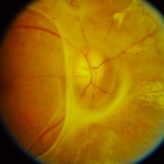
Ultra-wide field fundus photos of an 53-year-old man with a Subluxation of the Lens in the posterior vitreous cavity of the right eye after a trauma that happened many years ago. Patient remains stable with no adverse reaction to the lens at this time. No surgical intervention is recommended at this time. Patient also has myopic degeneration and lattice degeneration that will require patient to have follow up care.
Photographer: Kimberly Wakester, COA
Imaging device: Optos California
Condition/keywords: lattice degeneration, myopic degeneration, peripapillary atrophy, posterior staphyloma, Subluxation of the Lens
-
 Subluxation of the Lens
Dec 12 2024 by Kimberly Wakester
Ultra-wide field fundus photos of an 53-year-old man with a Subluxation of the Lens in the posterior vitreous cavity of the right eye after a trauma that happened many years ago. Patient remains stable with no adverse reaction to the lens at this time. No surgical intervention is recommended at this time. Patient also has myopic degeneration and lattice degeneration that will require patient to have follow up care.
Photographer: Kimberly Wakester, COA
Imaging device: Optos California
Condition/keywords: lattice degeneration, myopic degeneration, peripapillary atrophy, posterior staphyloma, Subluxation of the Lens
-
 Active Proliferative Diabetic Retinopathy
Jul 12 2024 by Korey Starkey
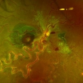
Fluorescein angiogram performed on 35 year old female with active proliferative diabetic retinopathy. Patient has peripapillary vascular loop and history of PRP treatment in both eyes. Patients left eye vision measured at Dcc20/200-1 at this visit.
Photographer: Korey Starkey
Imaging device: Optos
Condition/keywords: FLUORESCEIN ANGIOGRAPHY, hyperfluorescence, laser scarring, Optos, proliferative diabetic retinopathy (PDR), sea fan, ultra-wide field imaging, vascular loop
-
 Focal Chorioretinitis
Jul 11 2024 by Virginia Gebhart
67 year old female with punched-out CR scars. Hx of laser 3x for apparent peripapillary CNV. ESR, CRP, toxo, IgG/IgM all "normal." Bartonella, quant gold, and FTA-ABS ordered given possibility of neuroretinitis. Vision CF
Photographer: Virginia Gebhart
Imaging device: Optos California
Condition/keywords: FA, fluorescein angiogram (FA), FLUORESCEIN ANGIOGRAPHY, focal chorioretinitis, optic neuritis
-
 Pseudoxanthoma Elasticum with Angioid Streaks
May 10 2024 by Ethan K Sobol, MD
An asymptomatic patient with biopsy proven pseudoxanthoma elasticum. Both eyes had prominent peripapillary angioid streaks and a peau d'orange fundus appearance in the temporal macula.
Condition/keywords: angioid streaks, peau d'orange fundus, pseudoxanthoma elasticum (PXE)
-
 Serpiginous Choroidopathy
Apr 21 2024 by César Adrián Gómez Valdivia, MD
Gray-yellowish subretinal infiltrates that usually spread centrifugally from the peripapillary region in a serpiginous (snake-like) manner. Active lesions show a leading edge and resolve with subsequent RPE and choriocapillary atrophy.
Photographer: Erika Paulina Ornelas Cazares
Imaging device: TOPCON TRC-50DX
Condition/keywords: macula serpiginous choroidopathy
-
 Serpiginious Choroidopathy
Mar 21 2024 by Ogugua Ndubuisi Okonkwo, MD, FRCS (Edin), FASRS
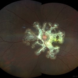
This is a left eye widefield fundus photograph of a 13-year-old male with a peripapillary ring of fibrotic scar that extends subretinally in finger-like projects along the vascular arcades and into the macula.
Photographer: Zainab Ogunsanu, Eye Foundation Hospital & Eye Foundation Retina Institute, Lagos.
Imaging device: ZEISS CLARUS 700
Condition/keywords: serpiginous like choroiditis
-
 Serpiginous Choroidopathy
Mar 21 2024 by Ogugua Ndubuisi Okonkwo, MD, FRCS (Edin), FASRS
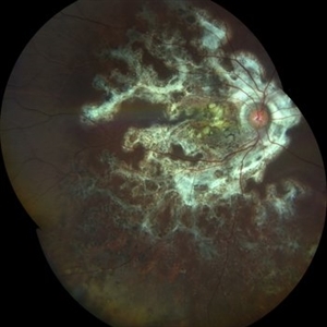
This is a right eye widefield fundus photograph of a 13-year-old male with a peripapillary ring of fibrotic scar that extends subretinally in finger-like projects along the vascular arcades and into the macula, with an extension of the scarring into the inferior retina, where it appears as a pigmented mottling.
Photographer: Zainab Ogunsanu, Eye Foundation Hospital & Eye Foundation Retina Institute, Lagos
Imaging device: ZEISS CLARUS 700
Condition/keywords: serpiginous like choroiditis
-
 Left Eye Arteriovenous Malformation, Vein Occlusion and Ruptured Macroaneurysm
Feb 9 2024 by Sandra R Montezuma, MD
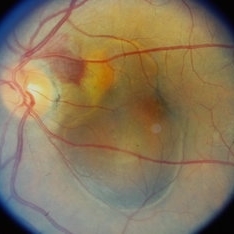
47 year old female presented with acute changes in vision in the left eye, flashes of light and a new supero temporal scotoma. No history of trauma. She has history of retina bleeding in 1998 when she was pregnant and had pre-eclampsia. She was told had a retina scar. Her VA was 20/500. Fundus exam revealed an arteriovenous malformation along inferonasal vessels with prominent tortuous vessels. The optic nerve was hyperemic and there was peripapillary pre-retinal hemorrhage. There is a central macula scar and retina hemorrhage in the macula and mid periphery. In the nasal mid periphery, there is a ruptured macroaneurysm with hemorrhage in all layers of the retina. There are diffuse IRH. Her OCT revealed abnormal foveal contour with intraretinal fluid, Outer retinal atrophy and increased hyperreflectivity of the inner retina layers. The patient was treated with avastin injections with some improvement of the vision and resolution of the intraretinal fluid. Her MRI was normal.
Photographer: University of Minnnesota
Condition/keywords: arteriovenous malformation, macroaneurysm, vein occlusion
-
 Multimodal Imaging for Differentiating Unilateral Pseudo Optic Disc Swelling(Buried Drusen) From True Optic Disc Swelling
Feb 7 2024 by Fawwaz F Al Mamoori, MD, Medical Retina Consultant
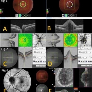
27-year-old male, medically free, presented with left unilateral optic disc swelling. BCVA=1.0(OU), color vision, and contrast sensitivity were normal (OU)with no RAPD in the left eye. Swept Source OCT: showed elevated left optic disc with hyporeflective mass (Fig-1 B). Enface OCT: Showed left peripapillary multiple ovoid mass lesions(drusen) (Fig-2 d, Fig3 F). FAF: of the left eye showed superonasal hyper autofluorescent drusenoid lesions)(Fig3 E). Orbital MRI with contrast was requested to exclude any compressive lesions like tumors(menigioma)or inflammatory lesions like granuloma(sarcoid granuloma). orbital MRI result was normal.
Photographer: Hana.S.Owais
Imaging device: TRITON(TOPCON,Swept Source OCT)
Condition/keywords: fundus autofluorescence (FAF), multimodal imaging, OCT EN FACE, optic disc drusen, optic disc edema, swept source
-
 Multimodal Imaging for Differentiating Unilateral Pseudo Optic Disc Swelling(Buried Drusen) From True Optic Disc Swelling
Feb 7 2024 by Fawwaz F Al Mamoori, MD, Medical Retina Consultant
A 27-year-old male patient, medically free, presented with unilateral left optic disc swelling. BCVA=1.0(OU), color vision, and contrast sensitivity were normal (OU) with no RAPD in the left eye. SS-OCT: showed left optic disc elevation with hyporeflective mass lesion (Fig-1 B). Enface OCT: showed left peripapillary hyperreflective ovoid mass lesions(Fig-2 D, Fig-3 F), FAF: showed left superonasal hyperautofluorescent drusenoid lesions. Orbital MRI with contrast was requested to exclude any optic nerve compressive lesions like (tumors: like mengioma or inflammatory lesions like granuloma (sarcoidosis). the result of orbital MRI was normal.
Photographer: Hana.S.Owais
Imaging device: TRITON(TOPCON,Swept Source OCT)
Condition/keywords: fundus autofluorescence (FAF), multimodal imaging, OCT EN FACE, optic disc drusen, optic disc edema
-
 Non invasive multimodal imaging for differentiating unilateral pseudo swelling buried optic disc drusen from true optic disc swelling
Feb 7 2024 by Fawwaz F Al Mamoori, MD, Medical Retina Consultant
27-year-old male, medically free, routine fundus examination showed left optic dic swelling, BCVA =1.0(OU), color vision, and contrast sensitivity were normal with no RAPD (OU). SS-OCT of the left optic disc showed a hyporeflective mass. Enface OCT shadogram showed peripapillary ovoid structures (drusen).FAF: showed drusenoid autofluorescence in the superonasal part only. Orbital MRI with contrast was requested to exclude any optic nerve tumor and it was normal.
Photographer: Hana.S.Owais
Imaging device: TRITON(OCT) Topcon
Condition/keywords: multimodal imaging, optic disc drusen, optic disc swelling
-
 Idiopathic Peripapillary CNV
Jan 4 2024 by Virginia Gebhart
13 year old female with inactive CNV. Increased pigment 360 at 1 year follow up. No inflammation or SRF, pt remains asymptomatic
Photographer: Virginia Gebhart
Imaging device: Optos California
Condition/keywords: choroidal neovascularization (CNV), peripapillary choroidal neovascularization (PPCNVM)
-
 Central Retinal Vein Occlusion associated with disc edema
Oct 19 2023 by Gabriel Costa Andrade, PhD
53-year-old woman with an acute CRVO. The patient has a history of breast cancer undergoing treatment with systemic chemotherapy. Notice the peripapillary cotton wool spots, superficial flame shaped hemorrhages and deeper dot and blot hemorrhages in all 4 quadrants.
Photographer: Gabriel Andrade
Condition/keywords: central retinal vein occlusion (CRVO), macular edema, Retina
-
 Peripapillary Choroidal Neovascular Membrane (CNVM)
Sep 26 2023 by Ben Serar
Fundus photograph showing Peripapillary Choroidal Neovascular Membrane (CNVM) with subretinal bleed and surrounding sub retinal fluid.
Condition/keywords: Peripapillary Choroidal Neovascular Membrane (CNVM)
-
 Peripapillary Choroidal Neovascular Membrane (CNVM) with Angioid Streaks
Sep 21 2023 by Ben Serar
Fundus photograph of RE showing Peripapillary Choroidal Neovascular Membrane (CNVM) nasal to disc with blurring of nasal disc margin and superficial retinal haemorrhages, with peripapillary angioid streaks.
Condition/keywords: angioid streaks, Peripapillary Choroidal Neovascular Membrane (CNVM)
-
 Peripapillary Atrophy
Sep 21 2023 by Ben Serar
Fundus photograph showing peripapillary atrophy.
Condition/keywords: peripapillary atrophy
-
 Peripapillary Choroidal Neovascular Membrane (CNVM)
Sep 21 2023 by Ben Serar
Fundus photograph of RE showing Peripapillary Choroidal Neovascular Membrane (CNVM) temporal to the disc with surrounding exudates.
Condition/keywords: Peripapillary Choroidal Neovascular Membrane (CNVM)
-
 Peripapillary traction
Sep 12 2023 by Ben Serar
Fundus photograph showing peripapillary traction , extending towards the vascular arcades.
Condition/keywords: traction
-
 Peripapillary Congenital Hypertrophy of the Retinal Pigment Epithelium
May 31 2023 by Luis Pimentel Silva, MD
Late phase Fluorescein Angiography of an 51 years old man with Peripapillary Congenital Hypertrophy of the Retinal Pigment Epithelium
Photographer: Luis Pimentel Silva, University of São Paulo
Condition/keywords: Peripapillary Congenital Hypertrophy
 Loading…