 Initializing download.
-
 By Fawwaz F Al Mamoori, MD, Medical Retina Consultant
Sharif Eye Center
Co-author(s): Dr.Omar Q - Uploaded on Feb 7, 2024.
- Last modified by Joshua Friedman on Feb 7, 2024.
- Rating
- Appears in
- Miscellaneous
- Condition/keywords
- optic disc drusen, optic disc swelling, multimodal imaging
- Photographer
- Hana.S.Owais
- Imaging device
-
Optical coherence tomography system
TRITON(OCT) Topcon - Description
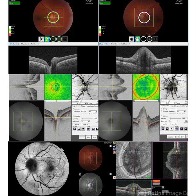
- 27-year-old male, medically free, routine fundus examination showed left optic dic swelling, BCVA =1.0(OU), color vision, and contrast sensitivity were normal with no RAPD (OU). SS-OCT of the left optic disc showed a hyporeflective mass. Enface OCT shadogram showed peripapillary ovoid structures (drusen).FAF: showed drusenoid autofluorescence in the superonasal part only. Orbital MRI with contrast was requested to exclude any optic nerve tumor and it was normal.


---thumb.jpg/image-square;max$79,0.ImageHandler "Optic Disc Drusen")
---thumb.jpg/image-square;max$79,0.ImageHandler "Optic Disc Drusen")


