Search results (569 results)
-
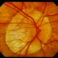
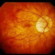
---thumb.jpg/image-square;max$300,300.ImageHandler) Presumed Ocular Histoplasmosis Syndrome
Presumed Ocular Histoplasmosis Syndrome
Feb 26 2013 by Henry J. Kaplan, MD
Color fundus photograph of the right eye of a patient with POHS shows typical punched out scars and peripapillary atrophy.
Condition/keywords: presumed ocular histoplasmosis syndrome (POHS)
-
---thumb.jpg/image-square;max$300,300.ImageHandler) Myopic fundus
Myopic fundus
Jan 11 2013 by Hyung-Woo Kwak, MD
Myopic fundus reveals yellow-colored lacquer cracks and peripapillary atrophy. There was visible choroidal vessel due to thin retina.
Photographer: Misook Lee, Kyung Hee Univsersity Hospital, Seoul
Imaging device: Zeiss f 450 plus
Condition/keywords: myopic fundus
-
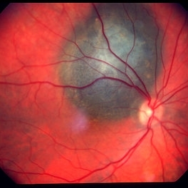
 Peripapillary Atrophy With High Myopia
Feb 4 2015 by H. Michael Lambert, MD
Peripapillary atrophy and central macular degeneration seen in high myopia.
Condition/keywords: high myopia, peripapillary atrophy
-
 Choroidal melanoma case 3 - peripapillary
Jan 11 2013 by Alex P. Hunyor, MD
Right peripapillary choroidal melanoma.
-
---thumb.jpg/image-square;max$300,300.ImageHandler) Peripapillary Atrophy
Peripapillary Atrophy
Feb 13 2013 by From the Collections of Thomas M. Aaberg, MD and Thomas M. Aaberg Jr., MD
Papilledema, intra-retinal hemorrhage, periopticneuritis.
Condition/keywords: intraretinal hemorrhage, papilledema, periopticneuritis, peripapillary atrophy
-
 Choroidal Melanoma
May 2 2013 by Henry J. Kaplan, MD
Peripapillary choroidal melanoma.
-
 Polypoidal Choroidal Vasculopathy
Aug 25 2012 by Hamid Ahmadieh, MD
FA & ICG angiography imagings of a 73-year-old man with a peripapillary PCV.
Photographer: Hamid Ahmadieh, Ophthalmic Research Center, Labbafinejad Medical Center
Imaging device: Heidelberg Spectralis
Condition/keywords: indocyanine green (ICG) angiography, polypoidal choroidal vasculopathy (PCV)
-
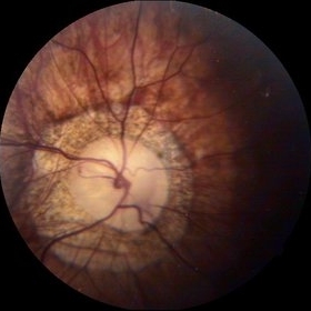
---thumb.jpg/image-square;max$300,300.ImageHandler) Presumed Ocular Histoplasmosis Syndrome
Presumed Ocular Histoplasmosis Syndrome
Feb 26 2013 by Henry J. Kaplan, MD
POHS; left eye: large punched out pigmented scars and peripapillary atrophy.
Condition/keywords: presumed ocular histoplasmosis syndrome (POHS)
-
 Superior Peripapillary Hemorrhage
Jul 13 2013 by Jason S. Calhoun
Patient was seen for acute vision loss in the right eye. Patient has glaucoma. VA was 20/70 in the right eye. Had vitrectomy back in May 2012 for ERM stripping. Also had trabectome with cataract surgery in December of 2012. Fundus photos presents a superior peripapillary Hemorrhage of the optic nerve. Patient will be followed up in one month.
Photographer: Jason S. Calhoun, Department of Ophthalmology, Mayo Clinic Jacksonville, Florida
Imaging device: TOPCON TRC 50-EX
Condition/keywords: peripapillary hemorrhage
-
 Peripapillary Atrophy With High Myopia
Feb 4 2015 by H. Michael Lambert, MD
Peripapillary atrophy and central macular degeneration seen in high myopia.
Condition/keywords: high myopia, peripapillary atrophy
-
---thumb.jpg/image-square;max$300,300.ImageHandler) Central Retinal Vein Occlusion
Central Retinal Vein Occlusion
Oct 30 2012 by Lihteh Wu, MD
35-year-old hypertensive man with an acute CRVO. Notice the peripapillary cotton wool spots, superficial flame shaped hemorrhages and deeper dot and blot hemorrhages in all 4 quadrants. This is the typical blood and thunder appearance of a CRVO.
Condition/keywords: central retinal vein occlusion (CRVO), cotton wool spots
-
---thumb.JPG/image-square;max$300,300.ImageHandler) Traumatic Optic Neuropathy
Traumatic Optic Neuropathy
Dec 9 2012 by Mallika Goyal, MD
Right eye of a 23-year-old gentleman 6 months following a road accident. Optic disc pallor with peripapillary chorioretinal scarring suggests traumatic optic neuropathy as the cause of optic atrophy.
Photographer: Mallika Goyal, MD, Apollo Health City, Hyderabad, India
Condition/keywords: traumatic optic neuropathy
-
 Peripapillary Atrophy
Oct 3 2014 by Mehul A Shah
A 55-year-old patient presented with diminished vision OU on examination patient had glaucoma with peripapillary optic atrophy.
Photographer: Drashti Netralaya,Dahod
Imaging device: Zeiss ff450
Condition/keywords: atrophy, peripapillary
-
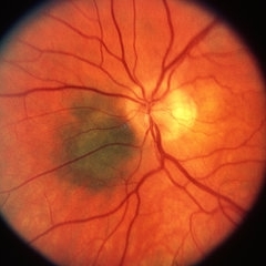
 Peri-Papillary CNVM
Oct 2 2013 by Jerald A. Bovino, MD
There is a peripapillary cnroidal neovascular membrane visible as the hyperfluorescent area temporal to the disk.
Condition/keywords: peripapillary
-
---thumb.jpg/image-square;max$300,300.ImageHandler) Tuberous Sclerosis
Tuberous Sclerosis
Feb 13 2013 by From the Collections of Thomas M. Aaberg, MD and Thomas M. Aaberg Jr., MD
Mulberry lesion.
Condition/keywords: peripapillary, tuberous sclerosis
-
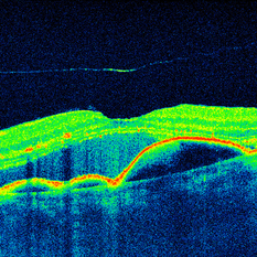
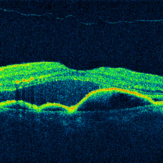 OCT Cirrus 5 Line Scan ARMD SRF RPED Stage 3 PVD
Mar 6 2013 by James B. Soque, CRA, OCT-C, COA, FOPS
Zeiss Cirrus OCT 4000, 5 Line Scan , 91-year-old white female with peripapillary SRN with subretinal heme, serous fluid, and a stage 3 PVD, still attached at the optic nerve.
Photographer: James Soque, CRA, COA, Island-Retina
Imaging device: Zeiss Cirrus 4000 SD OCT with 6.0.2.81 Software
Condition/keywords: optical coherence tomography (OCT)
-
 Buried drusen with CNV
Dec 19 2012 by Eric A. Postel, MD
Buried optic disc drusen complicated by peripapillary subretinal neovascularisation
Condition/keywords: choroidal neovascularization (CNV), optic disc drusen
-
 Traumatic Peripapillary Hemorrhage
May 2 2013 by Henry J. Kaplan, MD
Peripapillary subretinal hemorrhage in the left eye after blunt trauma.
Condition/keywords: blunt trauma, peripapillary hemorrhage, subretinal hemorrhage
-
---thumb.jpg/image-square;max$300,300.ImageHandler) Central Retinal Vein Occlusion
Central Retinal Vein Occlusion
Oct 30 2012 by Lihteh Wu, MD
35-year-old hypertensive man with an acute CRVO. Notice the peripapillary cotton wool spots, superficial flame shaped hemorrhages and deeper dot and blot hemorrhages in all 4 quadrants. This is the typical blood and thunder appearance of a CRVO.
Condition/keywords: central retinal vein occlusion (CRVO), cotton wool spots
-
 Peri-Papillary CNVM
Oct 2 2013 by Jerald A. Bovino, MD
There is a peripapillary cnroidal neovascular membrane visible as the yellow-white areas surrounded by hemorrhage temporal to the disk.
Condition/keywords: peripapillary
-
 Papilledema
Sep 21 2012 by Suber S. Huang, MD, MBA, FASRS
Fundus photograph of a 24-year-old obese woman with severe papilledema secondary to idiopathic intracranial hypertension.
Condition/keywords: dilated tortuous vessels, exudate, idiopathic intracranial hypertension, Paton's lines, peripapillary hemorrhage, pseudotumor cerebri
-
 OCT Cirrus 5 Line HD Scan EDI ARMD SRF RPED Stage 3 PVD
Mar 6 2013 by James B. Soque, CRA, OCT-C, COA, FOPS
Zeiss Cirrus OCT 4000, EDI Aquired Using Enhanced Depth Mode, 91-year-old white female with peripapillary SRN with subretinal heme, serous fluid, and a stage 3 PVD, still attached at the optic nerve.
Photographer: James Soque, CRA, COA, Island-Retina
Imaging device: Zeiss Cirrus 4000 SD OCT with 6.0.2.81 Software
Condition/keywords: optical coherence tomography (OCT)
-
 Peripapillary Glial Proliferation
Oct 18 2012 by Suber S. Huang, MD, MBA, FASRS
61-year-old woman with peripapillary gilal proliferation
Photographer: Stacie Hrvatin
Condition/keywords: glial proliferation, posterior vitreous detachment, Weiss ring
-
 Post Traumatic Optic Nerve Head Avulsion
Nov 18 2017 by Vishal Agrawal, MD, FRCS,FACS,FASRS
Right eye fundus picture of a 24-year-old male patient who suffered blunt trauma 7 days back with a wooden stick . He presented with NLP vision with a non reacting dilated pupil. Fundus montage picture shows ONH avulsion,CRAO,peripapillary resolving hemorrhages and cicatricial tissue at the edge.
Photographer: Vishal Agrawal, MD, SMS Medical College, Jaipur, India
Imaging device: Zeiss 524
Condition/keywords: avulsion, central retinal artery occlusion (CRAO)
-
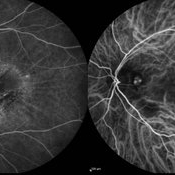
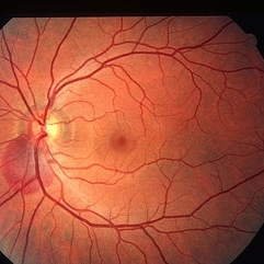
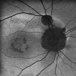 AZOOR
AZOOR
Mar 19 2015 by Niloofar Piri, MD
#1: Fundus autofluorescence OD in a patient with AZOOR demonstrates characteristic peripapillary hypoAF as well as concentric rings of hypo and hyper AF in posterior pole .
Imaging device: Heidelberg Spectralis
Condition/keywords: acute zonal occult outer retinopathy (AZOOR)
 Loading…