 Initializing download.
-
 By Fawwaz F Al Mamoori, MD, Medical Retina Consultant
Sharif Eye Center
Co-author(s): Dr.Omar Q - Uploaded on Feb 7, 2024.
- Last modified by Joshua Friedman on Feb 7, 2024.
- Rating
- Appears in
- Miscellaneous
- Condition/keywords
- optic disc drusen, optic disc edema, multimodal imaging, OCT EN FACE, fundus autofluorescence (FAF)
- Photographer
- Hana.S.Owais
- Imaging device
-
Optical coherence tomography system
TRITON(TOPCON,Swept Source OCT) - Description
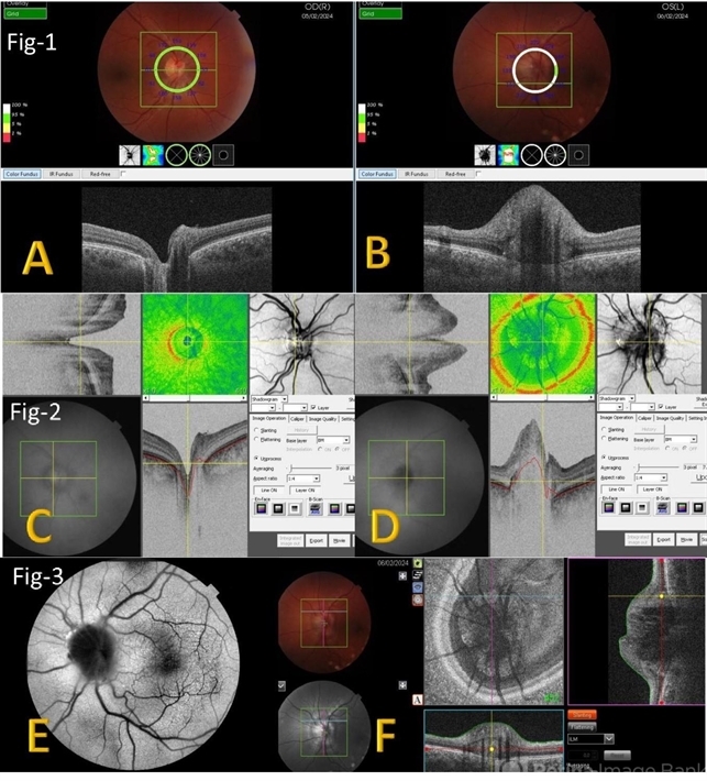
- A 27-year-old male patient, medically free, presented with unilateral left optic disc swelling. BCVA=1.0(OU), color vision, and contrast sensitivity were normal (OU) with no RAPD in the left eye. SS-OCT: showed left optic disc elevation with hyporeflective mass lesion (Fig-1 B). Enface OCT: showed left peripapillary hyperreflective ovoid mass lesions(Fig-2 D, Fig-3 F), FAF: showed left superonasal hyperautofluorescent drusenoid lesions. Orbital MRI with contrast was requested to exclude any optic nerve compressive lesions like (tumors: like mengioma or inflammatory lesions like granuloma (sarcoidosis). the result of orbital MRI was normal.


")


---thumb.jpg/image-square;max$79,0.ImageHandler "Polypoidal Choroidal Vasculopathy: Case 1 - Image 2 of 7")
