Search results (569 results)
-
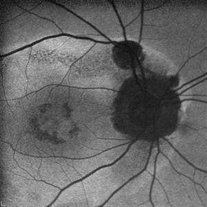
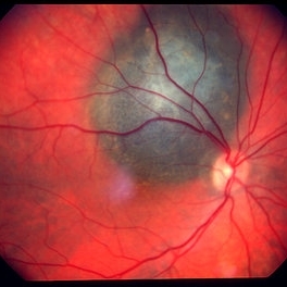
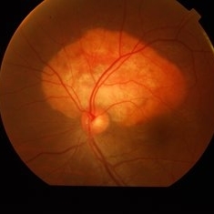
 Post Traumatic Optic Nerve Head Avulsion
Nov 18 2017 by Vishal Agrawal, MD, FRCS,FACS,FASRS
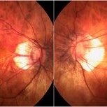
Right eye fundus picture of a 24-year-old male patient who suffered blunt trauma 7 days back with a wooden stick . He presented with NLP vision with a non reacting dilated pupil. Fundus montage picture shows ONH avulsion,CRAO,peripapillary resolving hemorrhages and cicatricial tissue at the edge.
Photographer: Vishal Agrawal, MD, SMS Medical College, Jaipur, India
Imaging device: Zeiss 524
Condition/keywords: avulsion, central retinal artery occlusion (CRAO)
-
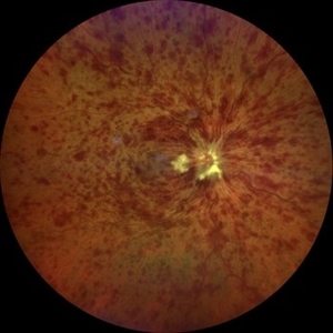
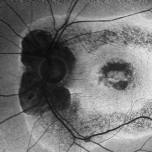
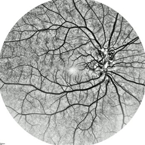
 AZOOR
AZOOR
Mar 19 2015 by Niloofar Piri, MD
#1: Fundus autofluorescence OD in a patient with AZOOR demonstrates characteristic peripapillary hypoAF as well as concentric rings of hypo and hyper AF in posterior pole .
Imaging device: Heidelberg Spectralis
Condition/keywords: acute zonal occult outer retinopathy (AZOOR)
-
---thumb.jpg/image-square;max$300,300.ImageHandler) Central Retinal Vein Occlusion
Central Retinal Vein Occlusion
Oct 30 2012 by Lihteh Wu, MD
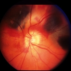
35-year-old hypertensive man with an acute CRVO. Notice the peripapillary cotton wool spots, superficial flame shaped hemorrhages and deeper dot and blot hemorrhages in all 4 quadrants. This is the typical blood and thunder appearance of a CRVO.
Condition/keywords: central retinal vein occlusion (CRVO), cotton wool spots
-
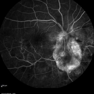
 Choroidal Granuloma
Apr 7 2017 by Manish Nagpal, MD, FRCS (UK), FASRS
Colour photo of a case of peripapillary choroidal granuloma presenting with exudation and hemorrhages.
Photographer: pooja barot
Condition/keywords: choroid, granuloma, inflammation
-
---thumb.jpg/image-square;max$300,300.ImageHandler) Central Retinal Vein Occlusion
Central Retinal Vein Occlusion
Oct 30 2012 by Lihteh Wu, MD
35-year-old hypertensive man with an acute CRVO. Notice the peripapillary cotton wool spots, superficial flame shaped hemorrhages and deeper dot and blot hemorrhages in all 4 quadrants. This is the typical blood and thunder appearance of a CRVO.
Condition/keywords: central retinal vein occlusion (CRVO), cotton wool spots
-
 Central Retinal Vein Occlusion associated with disc edema
Oct 19 2023 by Gabriel Costa Andrade, PhD
53-year-old woman with an acute CRVO. The patient has a history of breast cancer undergoing treatment with systemic chemotherapy. Notice the peripapillary cotton wool spots, superficial flame shaped hemorrhages and deeper dot and blot hemorrhages in all 4 quadrants.
Photographer: Gabriel Andrade
Condition/keywords: central retinal vein occlusion (CRVO), macular edema, Retina
-
 Choroidal Granuloma
Apr 7 2017 by Manish Nagpal, MD, FRCS (UK), FASRS
Fluorescein angiography of a case of peripapillary choroidal granuloma presenting with exudation and hemorrhages.
Photographer: Pooja Barot
Condition/keywords: choroid, granuloma, inflammation
-
 Choroidal melanoma case 3 - peripapillary
Jan 11 2013 by Alex P. Hunyor, MD
Right peripapillary choroidal melanoma.
-
 Commotio Retinae with Retinal Hemorrhages
Mar 27 2018 by Nichole Lewis
14-year-old male hit in the right eye with a stick. Commotio Retinae with retinal hemorrhages and peripapillary hemorrhage.
Photographer: Nichole Lewis
Condition/keywords: commotio retinae, peripapillary hemorrhage, retinal hemorrhage
-
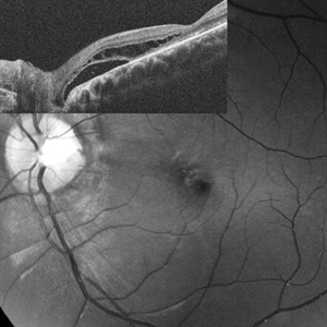 Optic Disc Pit Schisis RD
Apr 29 2013 by Michael Colucciello, MD, FASRS
Optic disc pit with peripapillary RD and macular schisis, fundus photograph and SD-OCT overlay.
Condition/keywords: optic disc pit
-
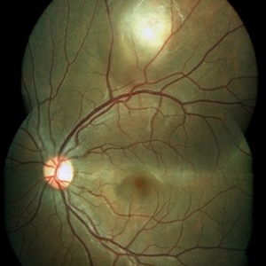
 Prepapillary Vascular Loop
Mar 11 2020 by Asdrubal F Moreno, MD
Fundus color photograph of a 80-year-old woman with a unilateral congenital prepapillary vascular loop and hypertensive retinopathy, focus on the vascular loop end for perception.
Photographer: Asdrubal Moreno, Fundacion AVAO, Universidad de Los Andes, Venezuela
Imaging device: Zeiss Visucam 500
Condition/keywords: congenital prepapillary vascular loop, peripapillary
-
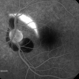
 AZOOR
AZOOR
Mar 19 2015 by Niloofar Piri, MD
#2 : Fundus autofluorescence OS in the same patient demonstrates more severe changes ; peripapillary hypoAF and concentric rings of hyper and hypo AF in posterior pole
Imaging device: Heidelberg Spectralis
Condition/keywords: acute zonal occult outer retinopathy (AZOOR)
-
 Choroidal rupture and peripapillary hemorrhage - FA
Jan 26 2013 by Roy Schwartz, MD
A 36-year-old male presented to the ER after blunt trauma to his left eye. On FA a chroidal rupture (hyperfluorescent area) was seen as well as peripapillary hemorrhage (hypofluorescent).
Photographer: Galit Yair-Pur
Condition/keywords: choroidal rupture, peripapillary hemorrhage
-
 High Myopia
Dec 7 2019 by Anfisa Ayalon, MD
Fundus photograph of a 55-year-old woman with high myopia.
Photographer: Anfisa Ayalon,MD., Meir Medical Center, Kfar Saba, Israel.
Condition/keywords: high myopia, myopia, peripapillary atrophy
-
 Idiopathic Peripapillary CNV
Jan 4 2024 by Virginia Gebhart
13 year old female with inactive CNV. Increased pigment 360 at 1 year follow up. No inflammation or SRF, pt remains asymptomatic
Photographer: Virginia Gebhart
Imaging device: Optos California
Condition/keywords: choroidal neovascularization (CNV), peripapillary choroidal neovascularization (PPCNVM)
-
 Maternally-Inherited Diabetes and Deafness (MIDD) Syndrome
Jan 12 2025 by Niloofar Piri, MD
Fundus Autofluorescence image of right posterior pole in a 43 year old female who was referred for diabetic retinopathy evaluation, demonstrated multiple patches of hypoautrofluorescence surrounding the nerve and fovea. Please note that central fovea is spared. Granular hyper and hypoauto fluorescence is present in the macula and peripapillary region. She was noted to have hearing loss as well and after further evaluation was diagnosed with MIDD syndrome.
Condition/keywords: Maternally inherited diabetes and deafness (MIDD), Maternally-inherited-diabetes-and-deafness-(MIDD) syndrome, Mitochondrial Disorder
-
 Peripapillary CNVM - ARMD
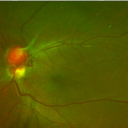
Jan 16 2014 by David Callanan, MD
Peripapillary CNVM - ARMD, 81-old-year female.
Condition/keywords: peripapillary
-
 Serpiginous Choroiditis With Peripapillary SRNVM
Apr 17 2017 by Manish Nagpal, MD, FRCS (UK), FASRS
patient having serpiginous choroiditis came with recent drop of central vision. Fundus revealed a streak of blood lining a membrane in the peripapillary lesion suggestive of a SRNVM.
Photographer: pooja barot
Condition/keywords: choroiditis, serpiginous choroiditis, subretinal neovascular membrane
-
 Retinal Capillary Hemangioma
Dec 12 2019 by David L Kilpatrick, MD
This is a wide-field color fundus photo showing two distinct retinal capillary hemangiomas. A visually significant epiretinal membrane is also present. Work up with gene testing was negative for VHL. The plan is to proceed with PDT of the two separate lesions (half fluence for the peripapillary lesion), followed by cryotherapy / photocoagulation.
Condition/keywords: retinal capillary hemangioma
-
---thumb.jpg/image-square;max$300,300.ImageHandler) Presumed Ocular Histoplasmosis Syndrome
Presumed Ocular Histoplasmosis Syndrome
Feb 26 2013 by Henry J. Kaplan, MD
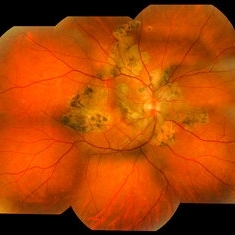
Color fundus photograph of the right eye of a patient with POHS shows typical punched out scars and peripapillary atrophy.
Condition/keywords: presumed ocular histoplasmosis syndrome (POHS)
-
 Prepapillary Vascular Loop
Mar 11 2020 by Asdrubal F Moreno, MD
Fundus color photograph of a 80-year-old woman with a unilateral congenital prepapillary vascular loop and hypertensive retinopathy, focused on the retinal plane for perception.
Photographer: Asdrubal Moreno, Fundacion AVAO, Universidad de Los Andes, Venezuela
Imaging device: Zeiss Visucam 500
Condition/keywords: congenital prepapillary vascular loop, peripapillary
-
 Choroidal Osteoma
Oct 11 2019 by Gayathri Mohan
Color fundus image of a peripapillary choroidal osteoma.
Photographer: Gayathri Mohan,Retina Foundation
Condition/keywords: choroidal osteoma, fundus photograph
-
---thumb.JPG/image-square;max$300,300.ImageHandler) Blastomycosis
Blastomycosis
Oct 28 2012 by Mallika Goyal, MD
Right eye fundus photograph of 32-year-old gentleman shows a large peripapillary choroidal granuloma. Lymph node biopsy had confirmed systemic blastomycosis. The choroidal granuloma resolved with systemic and intravitral voriconazole.
Condition/keywords: blastomycosis, choroidal granuloma
-
 Congenital Peripapillary Vascular Loops
Jun 22 2018 by Hashim Ali Khan, OD, FAAO
Inverted FA of a 10-year-old boy with congenital peripapillary vascular loop.
Imaging device: Spectraliz
Condition/keywords: congenital prepapillary vascular loop, pediatic retina
-
 Peripheral Choroidal Granuloma Associated With Tuberculosis Choroiditis
Jun 3 2017 by S. Natarajan, MD, FASRS, FRCS (GLASGOW) , FICO, D.Sc, FELA
Funds photograoh of an 21-year-old female pheripheral choroidal granuloma associated with tuberculos choroiditis.
Photographer: miss ashwini borde
Imaging device: Carl Zeiss 450 Plus IR
Condition/keywords: peripapillary choroidal granuloma
 Loading…