 Initializing download.
-
 By KANWALJEET HARJOT MADAN, M.S. (Ophthalmology); FAICO (Vitreous - Retina)
Thind Eye Hospital, Jalandhar City (Punjab). India. - Uploaded on May 4, 2025.
- Last modified by Joshua Friedman on May 5, 2025.
- Rating
- Appears in
- Retina Images
- Condition/keywords
- melanocytoma, optic nerve, melanoma
- Photographer
- Dr. Kanwaljeet Harjot Madan, Thind Eye Hospital, Jalandhar City (Punjab) INDIA.
- Imaging device
-
Fundus camera
Zeiss Fundus Camera - Description
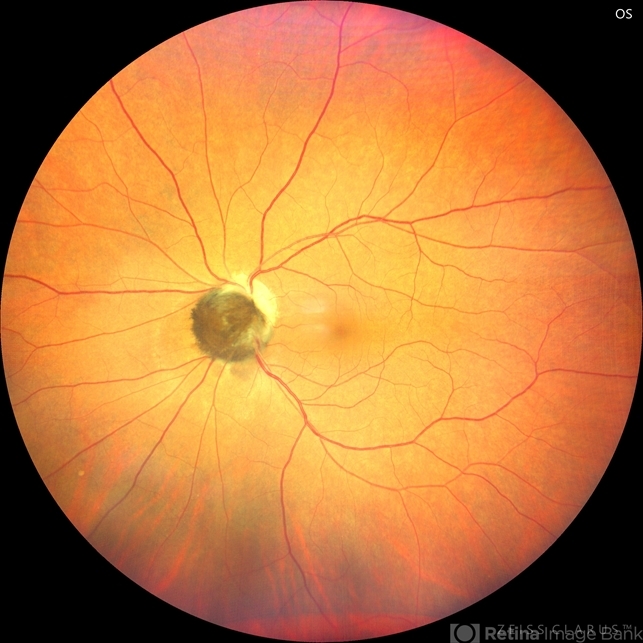
- This is a fundus picture of a young 42-year male who visited for a routine eye exam. His BCVA was 20/20 in both eyes. Anterior segment examination was normal. His left eye showed grey-black pigmentation at the infero-nasal margin of the optic disc. Fundus of the right eye was normal. The patient was diagnosed to have optic disc melanocytoma on multimodal imaging and was advised regular follow-up. Optic nerve melanocytoma is typically a benign tumor made up of melanocytes and melanin. It can grow, but rarely transforms into a malignancy. Patients with Optic Nerve Melanocytoma should be periodically examined for evidence of growth, loss of visual field and optic nerve compression.


