 Initializing download.
-
 By Tejaswita Verma
Retina Foundation hospital(Ahmedabad)
Co-author(s): DR. MANISH NAGPAL,RETINA FOUNDATION , AHMEDABAD - Uploaded on Sep 29, 2024.
- Last modified by Joshua Friedman on Sep 30, 2024.
- Rating
- Appears in
- Miscellaneous
- Condition/keywords
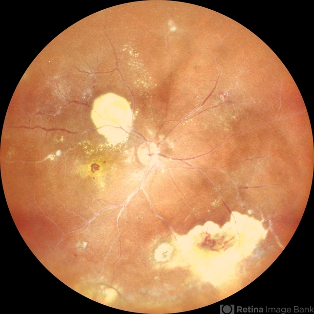
- Coats' disease
- Photographer
- DR. TEJASWITA VERMA
- Imaging device
-
Scanning laser ophthalmoscope
MIRANTE - Description
- Fundus photo of the RE of a 14 y/o female ,nil premorbid presented with reduced vision in the RE ,diagnosed incidentally on ophthalmological examination elsewhere .Vision was finger counting 3 meters in the RE . Fundus picture reveals macular scar , subretinal and intraretinal exudation ,with scattered hemorrhages esp. in STQ, sclerosed vessels in superior, superonasal quadrant ,nasal, inferonasal quadrant, CR scars inferiorly, Telengiectatic vessels S/O Coat's disease. She was advised RE anti VEGF x1 + laser PRP + PST kenacort under GA with guarded prognosis.


