Search results (1378 results)
-
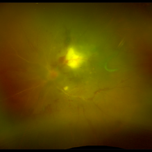
 Fluorescein Angiography in Choroidal Rupture
Jun 26 2025 by Hector Gabriel Moreno Solano, MD, MHA
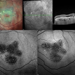
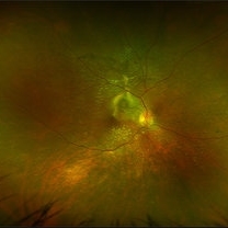
Fluorescein angiography of the right eye reveals three linear hypofluorescent lesions with progressive staining at the edges, consistent with choroidal ruptures. These lesions are temporally located in the posterior pole, with one of them situated near the fovea but without direct foveal involvement. The pattern is suggestive of previous blunt ocular trauma.
Photographer: Héctor Gabriel Moreno Solano, Instituto Mexicano de Oftalmología “IMO I.A.P”
Imaging device: CLARUS
Condition/keywords: Choroidal Rupture, fluorescein angiogram (FA)
-
 OCT Choroidal Rupture
Jun 26 2025 by Hector Gabriel Moreno Solano, MD, MHA
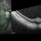
High-resolution OCT of the right eye shows a localized disruption of the retinal pigment epithelium (RPE)–Bruch’s membrane complex, consistent with a choroidal rupture. There is loss of the normal outer retinal architecture over the lesion, with focal elevation and irregularity of the underlying RPE. Hyperreflective material is noted at the level of the break, without associated subretinal fluid or signs of active choroidal neovascularization.
Photographer: Hector Gabriel Moreno Solano, Instituto Mexicano de Oftalmología “IMO I.A.P”
Imaging device: REVO
Condition/keywords: Choroidal Rupture, OCT
-
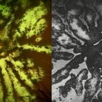
 Autofluorescence in Multiple Choroidal Ruptures
Jun 26 2025 by Hector Gabriel Moreno Solano, MD, MHA
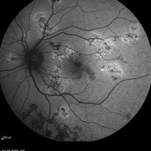
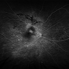
Fundus autofluorescence imaging of the right eye shows three hypoautofluorescent linear lesions located temporally to the fovea, consistent with choroidal ruptures. The lesions demonstrate sharply demarcated borders with variable surrounding hyperautofluorescence, suggestive of retinal pigment epithelium (RPE) disruption and potential remodeling. One rupture is located near the foveal region, though the foveal center remains spared.
Photographer: Hector Gabriel Moreno Solano, Instituto Mexicano de Oftalmología “IMO I.A.P”
Imaging device: CLARUS
Condition/keywords: autofluorescence imaging, Choroidal Rupture
-
 Multiple Chorodial Ruptures
Jun 26 2025 by Hector Gabriel Moreno Solano, MD, MHA
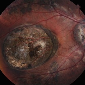
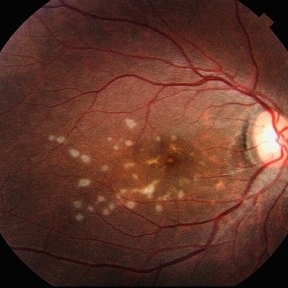
Color fundus photograph of the right eye reveals three well-defined, curvilinear choroidal ruptures located temporal to the fovea running parallel. The lesions appear as pale, crescent-shaped bands, with underlying retinal pigment epithelium disruption. One of the ruptures is situated near the foveal center, though without direct involvement.
Photographer: Hector Gabriel Moreno Solano, Instituto Mexicano de Oftalmología “IMO I.A.P”
Imaging device: CLARUS
Condition/keywords: Choroidal Rupture, color fundus photograph, color wide field
-
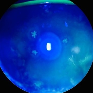 Dendritic Epithelial Keratitis
Jun 24 2025 by Arkaprava Ray
Slit-lamp photograph of a 46-year-old male showing dendritic epithelial lesion of herpes simplex keratitis, stained with 2% fluorescein sodium and viewed under cobalt blue filter.
Photographer: Arkaprava Ray, Trilochan Netralaya, Sambalpur, India
Condition/keywords: dendritic keratitis, HSV KERATITIS, keratitis
-
 RPE Rip s/p Brachytherapy for Malignant Melanoma
Jun 20 2025 by Virginia Gebhart
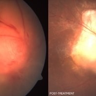
77 year old female with regressing tumor 4 months s/p brachytherapy. RPE rip at inferior edge of lesion.
Photographer: Virginia Gebhart, Retina Consultants of Carolina
Imaging device: Optos California
Condition/keywords: brachytherapy, choroidal melanoma, RPE Rip
-
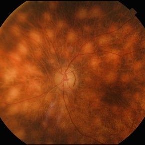 Birdshot Retinochoroidopathy
Jun 18 2025 by César Adrián Gómez Valdivia, MD
Fundus photograph of a 86 YO female patient diagnosed with Birdshot Retinochoroidopathy. Characteristically multifocal cream-colored or yellow-orange, oval or round lesions that emerge from around the optic nerve can be appreciated.
Photographer: @eyemissu2
Imaging device: TOPCON TRC-50DX
Condition/keywords: Birdshot Retinochoroidopathy
-
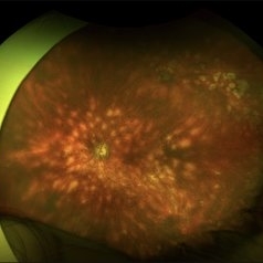 Birdshot Retinochoroidopathy
Jun 18 2025 by César Adrián Gómez Valdivia, MD
Fundus photograph of a 86 YO female patient diagnosed with Birdshot Retinochoroidopathy. Characteristically multifocal cream-colored or yellow-orange, oval or round lesions that emerge from around the optic nerve can be appreciated.
Photographer: @eyemissu2
Imaging device: California ICG OPTOS
Condition/keywords: Birdshot Retinochoroidopathy
-
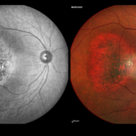 Choroidal Hemangioma
Jun 18 2025 by Moazzam Parvez
Multicolor and infrared reflectance image of a 42 year old gentleman with a Choroidal hemangioma lesion temporal to the fovea complaining of distortion in his right eye . Fundus imaging revealed a well-circumscribed ,elevated, reddish orange lesion within the choroid involving the posterior pole temporally .
Photographer: Moazzam Parvez, Netralayam , Kolkata
Imaging device: Heidelberg Spectralis
Condition/keywords: Choroidal Hemangioma, tumor
-
 Fractal Pattern of Chronic Serpiginous Choroiditis
Jun 17 2025 by Guilherme Sturzeneker, MD, MSc
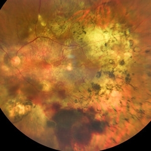
Ultra-widefield fundus photograph and autofluorescence of a 33-year-old woman with longstanding serpiginous choroiditis in the right eye. The image reveals centrifugal chorioretinal atrophy forming a dramatic fractal-like pattern, sparing the fovea. The patient is several years post-onset, with repeated negative workups, including for tuberculosis. Despite extensive lesions, the patient retains 20/20 vision in both eyes. Management included azathioprine monotherapy, as systemic steroids were contraindicated due to bipolar disorder.
Photographer: Andrea Almeida, IPEPO - Instituto da Visão
Imaging device: Optos Silverstone
Condition/keywords: autoimmune uveitis, azathioprine, chorioretinal atrophy, serpiginous choroiditis, ultra-wide field imaging
-
 The Headlight in the Fog
Jun 17 2025 by Thirumalesh Mochi Basavaraj, MD
37 year old male with sudden onset diminution of visual acuity has a large retinochoridal granuloma along the superotemporal arcade and a few with satellite lesions more temporal to it, there was extensive Occlusoive vasculitis (both arterioles and veins )being involved with Vitrities.
Photographer: Vivekanand ,Narayana nethralaya
Imaging device: Daytona
Condition/keywords: acute toxoplasmosis, retinochoroiditis
-
 Wagon-Wheel Lesion
Jun 5 2025 by César Adrián Gómez Valdivia, MD
Wagon-wheel lesion found in a 12 YO male patient diagnosed with congenital toxoplasmosis. Findings were bilateral.
Photographer: @eyemissu2
Imaging device: TOPCON TRC-50DX
Condition/keywords: toxoplasmosis chorioretinitis, Wagon-wheel lesion
-
 Wagon-Wheel Lesion
Jun 5 2025 by César Adrián Gómez Valdivia, MD
Wagon-wheel lesion found in a 12 year-old male patient diagnosed with congenital toxoplasmosis. Findings were bilateral.
Photographer: @eyemissu2
Imaging device: California ICG OPTOS
Condition/keywords: toxoplasmosis, Wagon-wheel lesion
-
 Wagon-Wheel Lesion
Jun 5 2025 by César Adrián Gómez Valdivia, MD
Wagon-wheel lesion found in a 12 year-old male patient diagnosed with congenital toxoplasmosis. Findings were bilateral.
Photographer: @eyemissu2
Imaging device: TOPCON TRC-50DX
Condition/keywords: toxoplasmosis, Wagon-wheel lesion
-
 Tuberculoma
Jun 4 2025 by Paulina Araujo
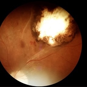
The 55-degree central fundus photograph of the left eye reveals an elevated, nodular whitish choroidal lesion along the inferior temporal arcade, consistent with a tuberculoma.
Photographer: Paulina D.Araujo Martínez, Asociación para Evitar la Ceguera en México I.A.P., Hospital Dr Luis Sánchez Bulnes.
Condition/keywords: Choroidal-tuberculoma
-
 Central Bouquet Hemorrhage
May 31 2025 by Moazzam Parvez
OCT image of a 26 year old gentleman of right eye macula with a central foveolar cotton ball like lesion . Inward traction by Müller cells over CB causes upward displacement of foveal cones without major disturbance of ellipsoid zone (EZ) and ELM . Cotton ball sign is characterized by small, fuzzy subfoveal hyperreflective area between the inner segment ellipsoid zone (EZ) and the interdigitation zone (IZ) .
Photographer: Dr Moazzam Parvez , Netralayam , Kolkata
Imaging device: Heidelberg Spectralis
Condition/keywords: Central bouquet haemorrhage, macula, myopia
-
 Multi-modal Imaging of Type - 1 CNVM
May 30 2025 by Shivankar Sen, MS, FVRS
Multimodal Imaging of a case of Polypoidal Choroidal Vasculopathy Multicolor Reflectance showing multiple green-hyper-fringent lesions in the macular region (Up Left) Infra-red Autofluorescence and Blue Autofluorescence showing hypo-autofluorescent areas correspondingly revealing the exact extent of the sub-RPE Lesion (Down left and right respectively) Optical Coherence Tomography - Enhanced Depth Imaging showing Thumb-shaped Pigment Epithelial Detachment with presence of Sub-retinal fluid and Hyper-reflective foci (Top Right)
Photographer: Dr. Shivankar Sen
Imaging device: Heidelberg Spectralis HRA+OCT
Condition/keywords: Blue autofluroscence, CNVM, multicolor, near infrared autofluorescence (NIRAF), PCV, reflectance
-
 Multiple evanescent White Dot Syndrome (MEWDS)
May 27 2025 by César Adrián Gómez Valdivia, MD
Fundus photograph of a 21 year-old female patient with suspected Multiple Evanescent White Dot Syndrome (MEWDS). The White Dot Syndromes produce yellow-white retinal lesions classically located at the retinal pigment epithelium or outer retina and are found primarily in young adults. Symptoms of MEWDS include unilateral blurred vision, visual field loss, photopsias, and floaters.
Photographer: @eyemissu2
Imaging device: TOPCON TRC-50DX
Condition/keywords: multiple evanescent white dot syndrome (MEWDS)
-
 Macular Star
May 27 2025 by César Adrián Gómez Valdivia, MD
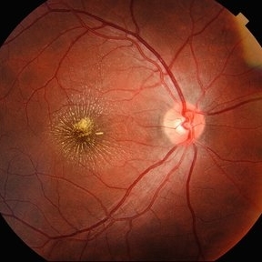
Macular Star found in a 31 year-old male patient with suspected Cat Scratch Disease. Typical intraocular presentations include neuroretinitis with optic nerve edema, macular star formation, and discrete white retinal or choroidal lesions. Findings were unilateral.
Photographer: @eyemissu2
Imaging device: TOPCON TRC-50DX
Condition/keywords: macular star
-
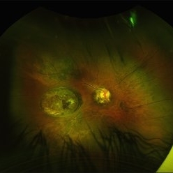
 Active multifocal choroiditis
May 26 2025 by Moazzam Parvez
Auto fluorescence photograph of an 43 year old man with active choroiditic lesion present in the left eye with recurrence
Photographer: Dr Moazzam Parvez , Netralayam , Kolkata
Imaging device: Heidelberg Spectralis
Condition/keywords: active choroididtis, choroiditi
-
 PCV
PCV
May 20 2025 by LUBNA AHMAD
Fundus image of a 66 year old male with huge branching vascular network, fresh hemorrhage and scarred PCV lesion.
Photographer: Sana Zamani
Imaging device: zeiss clarus 500
Condition/keywords: branching vascular network (BVN), polypoidal choroidal vasculopathy (PCV)
-
 VKH Pseudotumor – Chronic Subretinal Fibrosis
May 11 2025 by Felipe Murati
Ultra-widefield fundus image from a 36-year-old woman with chronic VKH syndrome showing a pseudotumor-like subretinal fibrotic lesion in the right eye. The lesion developed after multiple relapses and remained stable over a 1-year follow-up with immunosuppressive treatment including prednisone, mycophenolate mofetil, and adalimumab. No active choroiditis or exudative detachment was observed. Multimodal imaging was essential for disease monitoring.
Photographer: Felipe A. Murati, MD, University of Arizona
Imaging device: Optos California ultra-widefield retinal imaging system, single-capture, color fundus modality.
Condition/keywords: adalimumab, chronic inflammation, granulomatous uveitis, OCT, Optos ultra-widefield imaging, pseudotumor, subretinal fibrosis, VKH, Vogt-Koyanagi-Harada
-
 VKH Pseudotumor – Fluorescein Angiography
May 11 2025 by Felipe Murati
Fluorescein angiography image from a 36-year-old woman with chronic Vogt-Koyanagi-Harada (VKH) syndrome showing a pseudotumor-like lesion with late-phase staining and no active leakage. The image highlights subretinal fibrosis in the right eye, stable under long-term immunosuppressive therapy with mycophenolate mofetil and adalimumab. No signs of active choroiditis are present, confirming a quiescent phase.
Photographer: Felipe A. Murati, MD, University of Arizona
Imaging device: Optos California, fluorescein angiography modality
Condition/keywords: choroiditis, Fluorescein angiography, granulomatous uveitis, Optos FA, pseudotumor, subretinal fibrosis, VKH, Vogt-Koyanagi-Harada
-
 Optic Disc Granuloma
May 7 2025 by Aayesha - Khanum, MBBS. D.N.B
A 45-year-old male presented with diminished vision for one month. His Mantoux test was negative, but as steroids worsened the condition, quantiferon TB was advised and it returned positive. He was started on anti-tuberculosis treatment (ATT). Oral steroids were reintroduced after one week of ATT. Optic disc granulomas can arise from direct invasion of the optic nerve or may represent hypersensitivity reaction to tuberculous antigens. The pathogenesis involves infiltration of immune cells, leading to formation of a granulomatous structure that disrupts normal architecture and function of the optic disc. Steroids with ATT facilitated regression of granulomatous lesion.
Photographer: Ms. Krishna Jeyanthi
Imaging device: Zeiss Clarus 500
Condition/keywords: Tuberculosis
-
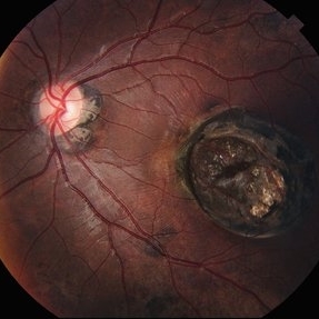
 Retinocoroiditis Inactiva Por Toxoplasmosis
Apr 28 2025 by Paulina Araujo
Fundus photography demonstrates a 2-disc-diameter chorioretinal scar in the superior temporal arcade, consistent with inactive toxoplasmic retinochoroiditis. The lesion exhibits pigmented borders and central atrophy, with adjacent splinter hemorrhages and vascular sheathing. No vitreous inflammation or active satellite lesions are present.
Photographer: Paulina D.Araujo Martínez, Asociación para Evitar la Ceguera en México I.A.P., Hospital Dr Luis Sánchez Bulnes.
Condition/keywords: toxoplasmosis chorioretinitis
 Loading…