 Initializing download.
-
 By Hector Gabriel Moreno Solano, MD, MHA
Instituto Mexicano de Oftalmología “IMO I.A.P!
Co-author(s): Claudia Gutiérrez Del Bosque, Instituto Mexicano de Oftalmología “IMO I.A.P” - Uploaded on Jun 26, 2025.
- Last modified by Joshua Friedman on Jun 30, 2025.
- Rating
- Appears in
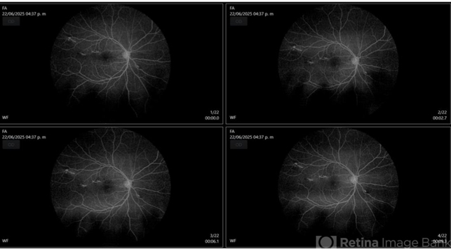
- Choroidal Rupture
- Condition/keywords
- Choroidal Rupture
- Photographer
- Héctor Gabriel Moreno Solano, Instituto Mexicano de Oftalmología “IMO I.A.P”
- Imaging device
-
Fundus camera
CLARUS - Description
- Fluorescein angiography of the right eye reveals three linear hypofluorescent lesions with progressive staining at the edges, consistent with choroidal ruptures. These lesions are temporally located in the posterior pole, with one of them situated near the fovea but without direct foveal involvement. The pattern is suggestive of previous blunt ocular trauma.


