 Daniel Davis, OCT-C
  Initializing download.
-
 By Guilherme Sturzeneker, MD, MSc
Co-author(s): Gabriela Yea-Huey Yang, MD; Rubens Belfort Neto, MD, PhD; Rubens Belfort Junior, MD, PhD, MBA - Uploaded on Jun 17, 2025.
- Last modified by Joshua Friedman on Jun 18, 2025.
- Rating
- Appears in
- Miscellaneous
- Condition/keywords
- serpiginous choroiditis, autoimmune uveitis, ultra-wide field imaging, azathioprine, chorioretinal atrophy
- Photographer
- Andrea Almeida, IPEPO - Instituto da Visão
- Imaging device
-
Scanning laser ophthalmoscope
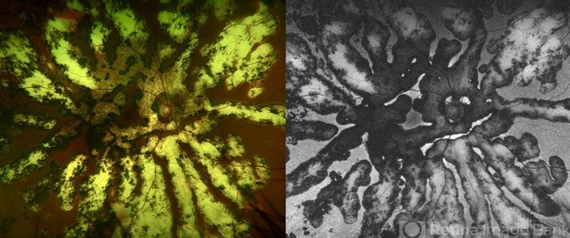
Optos Silverstone - Description
- Ultra-widefield fundus photograph and autofluorescence of a 33-year-old woman with longstanding serpiginous choroiditis in the right eye. The image reveals centrifugal chorioretinal atrophy forming a dramatic fractal-like pattern, sparing the fovea. The patient is several years post-onset, with repeated negative workups, including for tuberculosis. Despite extensive lesions, the patient retains 20/20 vision in both eyes. Management included azathioprine monotherapy, as systemic steroids were contraindicated due to bipolar disorder.


---thumb.JPG/image-square;max$79,0.ImageHandler "choroidal lymphoma")