Search results (1378 results)
-
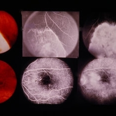
 Lattice Degeneration
Nov 9 2012 by Norman Byer
Lesion immediately adjacent to the ora serrata in an 18-year-old boy probably represents lattice degeneration characterized primarily by a reddish crater. It has remained unchanged for more than three years.
Condition/keywords: lattice degeneration, ora serrata, reddish crater
-
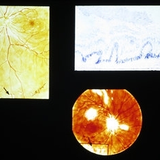
 Radiation
Feb 13 2014 by Howard Schatz, MD
Lesion (RB). Rad ret. Radiation.
Condition/keywords: radiation retinopathy
-
 Slide 8-25
Mar 4 2019 by Lancaster Course in Ophthalmology
Composite showing various lesions due to sarcoidosis. The drawings show seeding of sarcoid nodules in the vitreous (arrows). Histologic section through such a lesion shows granulomatous inflammatory aggregates extending into the vitreous from the retina (upper right).
Condition/keywords: lesions, sarcoid nodules, sarcoidosis
-
 'Headlight in the Fog' in Toxoplasmosis
Apr 8 2019 by Gary R. Cook, MD, FACS
29-year-old white male with moderate vitritis associated with an active toxoplasmosis lesion OS resulting in a "headlight in the fog" picture.
Condition/keywords: toxo chorioretinitis, toxoplasmosis, vitritis
-
 2:30 FA - Astrocytic Hemartoma
Oct 27 2019 by John S. King, MD
66-year-old white male without history of tuberous sclerosis was found to have an incidental, asymptomatic, translucent, retinal lesion with a few small telangiectatic vessels within it. The FA showed early hyperFL of these small vessels with prominent late leakage/staining. The OCT showed a retinal mass with a "moth eaten" appearance. Vision was 20/20 and the rest of the exam was unremarkable.
Photographer: Maisee Yang
Condition/keywords: astrocytic hamartoma
-
 28 Sec (Laminar Flow) FA - Astrocytic Hemartoma
Oct 27 2019 by John S. King, MD
66-year-old white male without history of tuberous sclerosis was found to have an incidental, asymptomatic, translucent, retinal lesion with a few small telangiectatic vessels within it. The FA showed early hyperFL of these small vessels with prominent late leakage/staining. The OCT showed a retinal mass with a "moth eaten" appearance. Vision was 20/20 and the rest of the exam was unremarkable.
Photographer: Maisee Yang
Condition/keywords: astrocytic hamartoma
-
 49 Sec FA - Astrocytic Hemartoma
Oct 27 2019 by John S. King, MD
66-year-old white male without history of tuberous sclerosis was found to have an incidental, asymptomatic, traslucent, retinal lesion with a few small telangiectatic vessels within it. The FA showed early hyperFL of these small vessels with prominent late leakage/staining. The OCT showed a retinal mass with a "moth eaten" appearance. Vision was 20/20 and the rest of the exam was unremarkable.
Photographer: Maisee Yang
Condition/keywords: astrocytic hamartoma
-
 61-Year-Old Man With Large Peripheral CHRPE
Dec 9 2017 by Timothy S Fuller, MD
61-year-old man presented for evaluation of pigmented retinal lesion. Found to have a large, peripheral CHRPE with characteristic lacunae, sharp margins, and lack of elevation.
Condition/keywords: benign pigmented lesions, congenital hypertrophy of the retinal pigment epithelium (CHRPE), lacunae
-
 A rare case of a 45-year-old male with choroidal neovascular membrane in Familial Dominant Drusen (Doyne Honeycomb Drusen) in both eyes treated with intravitreal injections.
Nov 30 2022 by SHRADDHA ASHOK CHANDORKAR, DNB DO
A 45-year-old man presented with diminution of vision in both eyes with metamorphopsia, which was painless and gradually progressive in nature. BCVA at presentation were 6/40 and 6/36 for the right and left eye respectively. Anterior segment examination of both eyes was unremarkable. IOP were within normal limits. Fundus examination showed bilateral numerous yellowish white round closely spaced lesions extending radially from the vascular arcades till the periphery associated with an elevated grayish macular choroidal neovascular membrane (CNV) with multiple drusen in the macular area and posterior pole. Impression was Familial Dominant Drusen (Doyne Honeycomb Drusen) associated with CNVM, both eyes. Color fundus photograph and autofluorescence showed Familial Dominant Drusen with CNVM. Subsequently , the patient underwent periodic intravitreal injections of Ranibizumab in both eyes under guarded visual prognosis, for which he tolerated well.
Photographer: NATIONAL INSTITUTE OF OPHTHALMOLOGY, PUNE
Imaging device: ZEISS CLARUS
Condition/keywords: choroidal neovascular membrane (CNVM), Doyne's Honeycomb, FAMILIAL DOMINANT DRUSEN, IMIM (Online Mendelian Inheritance in Man), intravitreal injection, Malattia Leventinese
-
---thumb.jpg/image-square;max$300,300.ImageHandler) Aborted Arteriolitis - diffuse hyper-permeability and staining of the infectious retinal lesion
Aborted Arteriolitis - diffuse hyper-permeability and staining of the infectious retinal lesion
Feb 15 2013 by From the Collections of Thomas M. Aaberg, MD and Thomas M. Aaberg Jr., MD
Fluorescein angiogram corresponding to slide titled Aborted Arteriolitis showing diffuse hyper-permeability and staining of the infectious retinal lesion.
Condition/keywords: ocular toxoplasmosis
-
 Active multifocal choroiditis
May 26 2025 by Moazzam Parvez
Auto fluorescence photograph of an 43 year old man with active choroiditic lesion present in the left eye with recurrence
Photographer: Dr Moazzam Parvez , Netralayam , Kolkata
Imaging device: Heidelberg Spectralis
Condition/keywords: active choroididtis, choroiditi
-
---thumb.jpg/image-square;max$300,300.ImageHandler) Active Toxoplasmosis
Active Toxoplasmosis
Aug 13 2013 by From the Collections of Thomas M. Aaberg, MD and Thomas M. Aaberg Jr., MD
Active lesion.
Condition/keywords: toxoplasmosis
-
---thumb.jpg/image-square;max$300,300.ImageHandler) Acute Toxoplasmosis
Acute Toxoplasmosis
Aug 13 2013 by From the Collections of Thomas M. Aaberg, MD and Thomas M. Aaberg Jr., MD
Active lesion.
Condition/keywords: toxoplasmosis
-
 Acute Exudative Polymorphous Vitelliform Maculopathy Angio OD
Aug 27 2014 by Flavio A. Rezende, MD, PhD
45-year-old man with mild decrease in vision after strong headache. Fundus showing multiple deep irregular vitelliform lesions spread throughout entire posterior pole OU, forming a typical level of subretinal confluent lesions at the inferior retinal vascular arcades. No primary tumor or metastasis found.
Photographer: Eduardo Martins, Pontifícia Universidade Católica - Rio de Janeiro, Brazil
Imaging device: Topcon TRC 50EX
Condition/keywords: polymorphous exudative vitelliform maculopathy
-
 Acute Exudative Polymorphous Vitelliform Maculopathy Angio OS
Aug 27 2014 by Flavio A. Rezende, MD, PhD
45-year-old man with mild decrease in vision after strong headache. Fundus showing multiple deep irregular vitelliform lesions spread throughout entire posterior pole OU, forming a typical level of subretinal confluent lesions at the inferior retinal vascular arcades. No primary tumor or metastasis found.
Photographer: Eduardo Martins, Pontifícia Universidade Católica - Rio de Janeiro, Brazil
Imaging device: Topcon TRC 50EX
Condition/keywords: polymorphous exudative vitelliform maculopathy
-
 Acute Exudative Polymorphous Vitelliform Maculopathy Color OD
Aug 27 2014 by Flavio A. Rezende, MD, PhD
45-year-old man with mild decrease in vision after strong headache. Fundus showing multiple deep irregular vitelliform lesions spread throughout entire posterior pole OU, forming a typical level of subretinal confluent lesions at the inferior retinal vascular arcades. No primary tumor or metastasis found.
Photographer: Eduardo Martins, Pontifícia Universidade Católica - Rio de Janeiro, Brazil
Imaging device: Topcon TRC 50EX
Condition/keywords: polymorphous exudative vitelliform maculopathy
-
 Acute Exudative Polymorphous Vitelliform Maculopathy Color OS
Aug 27 2014 by Flavio A. Rezende, MD, PhD
45-year-old man with mild decrease in vision after strong headache. Fundus showing multiple deep irregular vitelliform lesions spread throughout entire posterior pole OU, forming a typical level of subretinal confluent lesions at the inferior retinal vascular arcades. No primary tumor or metastasis found.
Photographer: Eduardo Martins, Pontifícia Universidade Católica - Rio de Janeiro, Brazil
Imaging device: Topcon TRC 50EX
Condition/keywords: polymorphous exudative vitelliform maculopathy
-
 Acute Exudative Polymorphous Vitelliform Maculopathy Red Free OD
Aug 27 2014 by Flavio A. Rezende, MD, PhD
45-year-old man with mild decrease in vision after strong headache. Fundus showing multiple deep irregular vitelliform lesions spread throughout entire posterior pole OU, forming a typical level of subretinal confluent lesions at the inferior retinal vascular arcades. No primary tumor or metastasis found.
Photographer: Eduardo Martins, Pontifícia Universidade Católica - Rio de Janeiro, Brazil
Imaging device: Topcon TRC 50EX
Condition/keywords: polymorphous exudative vitelliform maculopathy
-
 Acute Exudative Polymorphous Vitelliform Maculopathy Red Free OS
Aug 27 2014 by Flavio A. Rezende, MD, PhD
45-year-old man with mild decrease in vision after strong headache. Fundus showing multiple deep irregular vitelliform lesions spread throughout entire posterior pole OU, forming a typical level of subretinal confluent lesions at the inferior retinal vascular arcades. No primary tumor or metastasis found.
Photographer: Eduardo Martins, Pontifícia Universidade Católica - Rio de Janeiro, Brazil
Imaging device: Topcon TRC 50EX
Condition/keywords: polymorphous exudative vitelliform maculopathy
-
---thumb.jpg/image-square;max$300,300.ImageHandler) Acute Histoplasmosis Choroiditis In Immunocompetent Boy
Acute Histoplasmosis Choroiditis In Immunocompetent Boy
Oct 4 2013 by Maurice F. Rabb
On examination both brothers had visual acuities of 20/20 in each eye. The younger brother had a single distinct creamy-white 500 micron lesion located in the choroid temporal to the macula of the right eye. Fundus photographs of the older brother are shown. The right eye had at least five whitish lesions that appeared to be located deep to the retina. There were two lesions present superotemporal to the fovea, one immediately inferior to the fovea, one inferior to the optic nerve head, and one superonasally. In the left eye there was a single choroiditis lesion immediately nasal to the optic nerve head as well as some questionable lesions approximately one disc diameter inferior to the optic nerve head.
Condition/keywords: acute histoplasmosis choroiditis in immunocompetent boy
-
 Acute Histoplasmosis Choroiditis In Immunocompetent Boy
Oct 4 2013 by Maurice F. Rabb
On examination both brothers had visual acuities of 20/20 in each eye. The younger brother had a single distinct creamy-white 500 micron lesion located in the choroid temporal to the macula of the right eye. Fundus photographs of the older brother are shown. The right eye had at least five whitish lesions that appeared to be located deep to the retina. There were two lesions present superotemporal to the fovea, one immediately inferior to the fovea, one inferior to the optic nerve head, and one superonasally. In the left eye there was a single choroiditis lesion immediately nasal to the optic nerve head as well as some questionable lesions approximately one disc diameter inferior to the optic nerve head.
Condition/keywords: acute histoplasmosis choroiditis in immunocompetent boy
-
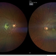
 Acute Macular Neuroretinopathy
Apr 12 2021 by Iuri Golubev, MD
46-year-old female with sudden onset paracentral scotoma below the central point of fixation in her left eye. Enface image shows a wedge shaped lesion pointing towards the fovea (top left). The lesion was spanning outer retinal layers from OPL to RPE (top left insert). One month later, the lesion has diminished in size, and was only involving retinal layers from ellipsoid zone to RPE(top right). At 4 months since presentation, the patient did not have any signs of AMN identifiable on enface or b-scan images (bottom center). Patient's symptoms has slowly improved and eventually resolved over the course of the next 4 years.
Imaging device: Zeiss Cirrus 5000
Condition/keywords: acute macular neuroretinopathy, acute macular outer retinopathy
-
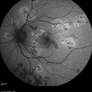
 Acute Macular Neuroretinopathy
Mar 25 2024 by Daniel Davis, OCT-C
18 yo female presenting with hazy vison for 2-3 weeks. VA OD: sc20/20 VA OS: sc20/20 Infrared imaging showed dark gray, petalloid, perifoveal lesions and OCT shows focal signal reduction of the Inner Segment / Outer Segment junction. Elects to observe.
Photographer: Daniel Davis, OCT-C, The Retina Institute, St. Louis
Imaging device: Optos California SWL
Condition/keywords: acute macular neuroretinopathy
-
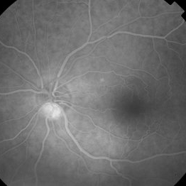
 Acute Macular Neuroretinopathy
Sep 15 2014 by Thomas A. Ciulla, MD, MBA, FASRS
Color photo. This might be a typical fundus photo, with no definite lesion. However, the infrared photo nicely depicts a typical lesion.
Condition/keywords: acute macular neuroretinopathy, color photo
 Loading…