 Initializing download.
-
 By David C Sousa, MD PhD FRANZCO
Co-author(s): Joshua Taylor MD & Angus Turner MBBS MSc FRANZCO, Lions Outback Vision, Lions Eye Institute, Nedlands, Western Australia, Australia - Uploaded on May 16, 2022.
- Last modified by Joshua Friedman on May 17, 2022.
- Rating
- Appears in
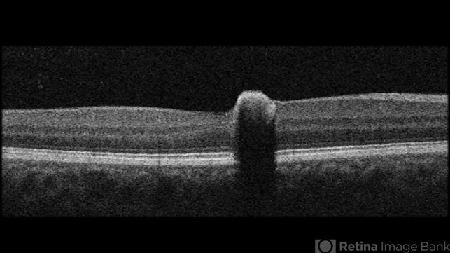
- Congenital Simple Hamartoma of the Retinal Pigment Epithelium
- Condition/keywords
- retinal pigment epithelium (RPE) hamartoma
- Imaging device
-
Optical coherence tomography system
Topcon Maestro2 - Description
- A 49-year-old man was referred after an incidental finding in the right eye macula. Best-corrected visual acuity was 20/25. Anterior segment examination was unremarkable. Fundoscopy revealed a juxta-foveal heavily pigmented well-demarcated slightly elevated lesion measuring 0.4 x 0.4 mm. No other changes were observed adjacent to the lesion or elsewhere in either eye. Optical coherence tomography revealed an area of retinal elevation with high optical reflectivity and posterior shadowing. The findings are consistent with congenital simple hamartoma of the retinal pigment epithelium. Given the benign, non-progressive and usually asymptomatic nature of this condition, most patients are diagnosed in adulthood.


