 Initializing download.
-
 By CUI YUELING
Co-author(s): Wei Tan,Xiaocong Wang - Uploaded on Jun 10, 2025.
- Last modified by Joshua Friedman on Jun 10, 2025.
- Rating
- Appears in
- Miscellaneous
- Condition/keywords
- commotio retinae
- Photographer
- Yueling Cui
- Imaging device
-
Fundus camera
Zeiss Clarus 500 - Description
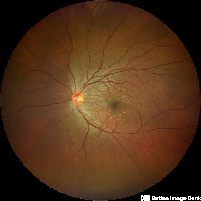
- The patient presented 2 hours after sustaining a left eye injury caused by a stick. Visual acuity in the left eye was 0.2 without improvement upon correction, and intraocular pressure measured 15 mmHg. Examination of the anterior segment revealed ciliary conjunctival injection accompanied by patchy subconjunctival hemorrhage. The corneal surface remained smooth, and the anterior chamber was deep with hyphema characterized by blood-tinged aqueous humor predominantly settled inferiorly. The pupil was slightly irregular, approximately 3 mm in diameter, with a superotemporal notch; pupillary light reflex was intact. The lens appeared clear. Fundus examination showed well-defined optic disc margins with normal coloration and a cup-to-disc ratio of 0.2. Retinal arteries and veins were normally distributed with an artery-to-vein ratio of 2:3. At the posterior pole, the foveal reflex exhibited concentric ripple-like changes centered on the fovea, accompanied by localized pigment attenuation and reduced reflex intensity. Irregular reflectivity was noted in the superotemporal and inferotemporal nerve fiber layers.

---thumb.jpg/image-square;max$79,0.ImageHandler "Severe Commotio Retinae With Edema And Hemorrhages")
---thumb.jpg/image-square;max$79,0.ImageHandler "Severe Commotio Retinae With Edema And Hemorrhages")
---thumb.jpg/image-square;max$79,0.ImageHandler "Severe Commotio Retinae With Edema And Hemorrhages")


