Search results (40 results)
-
 Sclerochoroidal Calcification
Apr 24 2025 by Virginia Gebhart
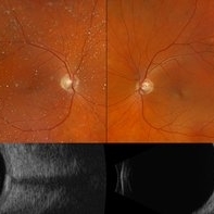
70 year old male referred for amelanotic lesion in the STA OU. Ultrasound shows slightly elevated lesions with hyperreflectivity and posterior shadowing with reduplication artifact consistent with sclerochoroidal calcification. Recommend yearly observation.
Photographer: Virginia Gebhart, Retina Consultants of Carolina
Imaging device: Optos California, Ellex Eye Cubed
Condition/keywords: asteroid hyalosis, B scan ultrasound, sclerochoroidal calcification
-
 IOL Reverberation
Apr 3 2025 by Gustavo Uriel Fonseca Aguirre
Top: Axial B-scan ultrasonography demonstrating an IOL with associated vitreous reverberation artifacts. Bottom: A-mode tracing revealing characteristic spike patterns consistent with IOL-induced reverberation.
Photographer: Gustavo U. Fonseca Aguirre, Hospital Conde de Valenciana, Ciudad de México
Condition/keywords: IOL, ultrasound
-
 Fundus Photo
Jun 6 2023 by Mayor Vang
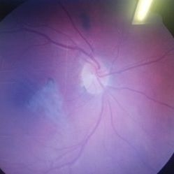
In honor of pride month! A fundus photo using a Ziess fundus camera showing an IOL artifact.
Photographer: Mayor Vang, (OCT-C, COT), UTSW, Dallas, TX
Condition/keywords: artifact, fundus photograph, IOL, optic disc
-
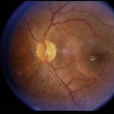
 Nevus
Nevus
Jan 21 2021 by AGNES KIM
Fundus photograph of 30-year-old female of choroidal nevus. Another nevus was found in the same eye in the periphery. Macula has a lens artifact.
Condition/keywords: choroidal nevus
-
 Ectopia Lentis
Jan 21 2021 by Jamin S. Brown, MD
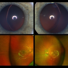
This image serial demonstrates a patient with simple ectopia lentis. Anterior segment photographs in the upper panel demonstrate nasally subluxated crystalline lenses. Widefield fundus photography shows a "pseudo-buckle" which is the result of an optical effect due to the lens subluxation (artifactual image enlargement). Also note the juvenile macular reflex in this young patient. Ectopia lentis can present isolated ("simple") or in combination with various systemic defects (Marfan's syndrome, Weil-Marchesani syndrome or Ehlers-Danlos syndrome to name a few). Isolated ectopia lentis can be hereditary and causative genes have been identified as ADAMTSL4 located on chromosome 4 and FBN1 gene located on chromosome 15. Defects in the genes cause weakness in the zonular fibers which can lead to lens dislocation. Lastly, various ocular disorders such as Aniridia, Axenfeld-Rieger, Pseudoexfoliation or Trauma may also result in lens dislocation or subluxation.
Photographer: Stefanie Palmer CRA, Retina Vitreous Surgeons of CNY
Condition/keywords: dislocated lens, ectopia lentis
-
 Puzzle Retinitis
Jan 20 2021 by Jamin S. Brown, MD
Puzzle artifact after imaging on a smaller field of view with blue light autofluorescence.
Photographer: Stefanie Palmer CRA, Retina Vitreous Surgeons of CNY
Condition/keywords: autofluorescence imaging, normal eye
-
 Retinoschisis with Outer Layer Break
Nov 4 2020 by Thomas A. Ciulla, MD, MBA, FASRS
Inferior temporal retinoschisis extending posteriorly to arcade. The vertical cut shows more severe splitting inferiorly, with visibly attached outer layer. The “inverted V” shape of the inner layer on the left of the image is artifact, due to OCT-algorithm related inversion as this layer extends towards the vitreous.
Condition/keywords: bullous retinoschisis, inferotemporal retinoschisis, outer layer breaks
-
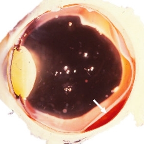
 Amelanotic Mushroom-Shaped Choroidal Melanoma
May 18 2020 by McGill University Health Centre
The enucleation specimen in (B) shows an amelanotic, mushroom-shaped, slightly hemorrhagic tumor near the optic nerve (arrow). The shape is due to infiltration of the retina by a rupture of the Bruch membrane. A retinal detachment artifact is present.
Condition/keywords: enucleation, mushroom-shaped
-
 Pigmented Tumor Infiltrating Ciliary Body
May 18 2020 by McGill University Health Centre
This enucleation specimen shows a heavily pigmented, solid tumor-infiltrating the ciliary body. The tumor infiltrates the left angle, producing secondary glaucoma. Note the dislocation of the lens and the choroidal detachment artifact on the right bottom angle of the image.
Condition/keywords: enucleation, infiltrating ciliary body, tumor
-
 Ciliary Body Melanoma
May 18 2020 by McGill University Health Centre
Large tumors displace the lens. Of the 3 locations in the uveal tract, tumors of the ciliary body have the worst prognosis. This enucleation specimen shows a pigmented, bilobed, dome-shaped tumor arising from the ciliary body (arrow). The lens has been removed, and a diffuse, flat retinal detachment artifact is present.
Condition/keywords: melanoma
-
 Ciliary Body Melanoma
May 18 2020 by McGill University Health Centre
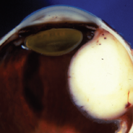
Uveal melanoma is the most common primary eye malignancy in adulthood, occurring mainly after age 60. The uveal tract — composed of the iris, ciliary body, and choroid — can be affected by uveal melanoma. Despite advances in treatment of the primary tumor, metastatic disease occurs in almost half of patients, generally affecting the liver and lungs via hematogenous dissemination of the primary tumor. Tumors have different levels of pigmentation, and some are amelanocytic (nonpigmented). The differential diagnosis for amelanotic choroidal melanoma is metastatic disease. Large tumors displace the lens. Of the 3 locations in the uveal tract, tumors of the ciliary body have the worst prognosis. The enucleation specimen in (A) shows a firm, dome-shaped, deeply pigmented tumor arising from the ciliary body (arrow). The lens has been removed, and a diffuse retinal detachment artifact is present.
Condition/keywords: enucleation, melanoma
-
 Mixed Retinoblastoma
May 18 2020 by McGill University Health Centre
This enucleation specimen shows a whitish, thickened area of the neurosensory retina over the head of the optic nerve, corresponding to a mixed retinoblastoma. Snowballlike structures are present in the vitreous chamber overlying the tumor, corresponding to vitreous seeding. A retinal detachment artifact is present.
Condition/keywords: mixed, retinoblastoma
-
 Metastatic Adenocarcinoma
May 18 2020 by McGill University Health Centre
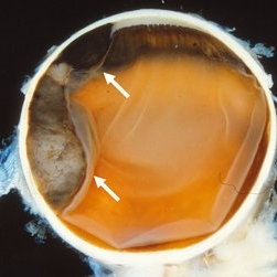
Metastatic disease is the most frequent intraocular malignant tumor. In women, the most common origin is breast cancer. In men, the most common origin is lung cancer. This pupil–optic nerve section shows a whitish tumor with several foci of necrosis (*) occupying the posterior aspect of the choroid. Note the pigment epithelium over the inner surface of the tumor. A serous retinal detachment is present (arrow) with a retinal detachment artifact overlying the tumor and normal choroid. Note the air bubble artifacts in the vitreous cavity. Another artifact, the compression of the eyeball, is present on the right side.
Condition/keywords: breast cancer, foci of necrosis, metastatic adenocarcinoma, tumor
-
 Leiomyoma
May 18 2020 by McGill University Health Centre
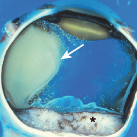
Leiomyoma is a benign, smooth muscle tumor. Ninety percent of cases occur in women. The differential diagnosis includes amelanotic melanoma and nerve sheath tumors. This transversal pupil–optic nerve (PO) section of an enucleation specimen shows a nodular, well-delineated, whitish tumor in the ciliary body. The cut surface shows small foci of hemorrhage without necrosis. The retina partially covers the inner surface of the tumor, and the sclera is not infiltrated. Note the slightly displaced (subluxated) cataractous lens and the choroidal detachment artifact in the right inferior corner.
Condition/keywords: enucleation, leiomyoma, tumor
-
 Enucleated Eye with Subretinal Hematoma
May 18 2020 by McGill University Health Centre
This enucleation specimen shows a subretinal hematoma (arrow). In addition, a diffuse, flat retinal detachment artifact is present. The lens is cataractous.
Condition/keywords: cataract, subretinal hemorrhage
-
 Common Artifacts in Macroscopic Ocular Globe Evaluation
May 18 2020 by McGill University Health Centre
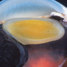
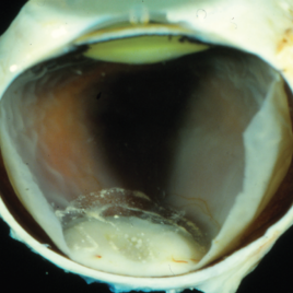
This sample was retrieved from a patient with a blind, painful eye. Blind, painful eye may be the end stage of several conditions including glaucoma, retinal detachment, and endophthalmitis, among others. Evisceration specimens are generally submitted in fragments. Different intraocular structures are identifiable: retina, cornea and capsular bag, choroidal tissue, and hematic material.
Condition/keywords: enucleation, evisceration, intraocular structures
-
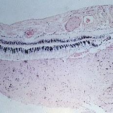 Slide 11-12
Feb 26 2019 by Lancaster Course in Ophthalmology
Myelin artifact ( x16). Myelin has been squeezed from nerve into retinal vessels and subretinal space, and may be confused with papilledema. (Scheie Eye Institute, No. 5031.)
Condition/keywords: myelin
-
 Optos Picture With Speculum: Dislocated Natural Lens
Oct 9 2018 by John S. King, MD
55-year-old white female with history of pathologic myopia+, lattice (laser), SB OU (1990s), and dislocated natural lenses OU that had been watched for years. In the fellow eye she developed phacolytic glaucoma and a PPV, PPL was performed. Plan for both eyes are monitoring. I wanted to get a good picture of her lens today with the optos machine, as the other pics had artifact from the lower lid. It worked out well to use a speculum in the left eye. Vision cc is 20/400 J1+ OD and 20/40 J2 OS; aphakic OU; vitreous clear OD; dislocated lens OS (see pic); retinas attached.
Photographer: Maisee Yang
Imaging device: Optos California
Condition/keywords: dislocated crystalline lens, pathologic myopia, scleral buckle, staphyloma
-
 Polypoidal Choroidal Vasculopathy
Dec 27 2014 by Thomas A. Ciulla, MD, MBA, FASRS
OCT revealed dome like elevation nasal to the foveal with surrounding subretinal fluid. The map shows thickening at the lesion with some artifact.
Condition/keywords: idiopathic polypoidal choroidal vasculopathy, polypoidal choroidal vasculopathy (PCV)
-
 RP Variant
Dec 22 2014 by H. Michael Lambert, MD
Fundus photo with lots of artifact.
Condition/keywords: RP variant
-
 Ultrasound Artifact From Intraocular Gas
Jul 9 2014 by Susanna S. Park, MD, PhD
B-scan ultrasonography showing reverberation artifacts from intraocular gas bubble filling 30% of the vitreous cavity and retinal detachment repair.
Photographer: Ellen Redenbo
Condition/keywords: intraocular gas, ultrasound
-
---thumb.jpg/image-square;max$300,300.ImageHandler) Series of Photos Demonstrating Photography Artifact
Series of Photos Demonstrating Photography Artifact
Feb 20 2013 by From the Collections of Thomas M. Aaberg, MD and Thomas M. Aaberg Jr., MD
FA.
Condition/keywords: artifact, camera, photography
-
---thumb.jpg/image-square;max$300,300.ImageHandler) Series of Photos Demonstrating Photography Artifact
Series of Photos Demonstrating Photography Artifact
Feb 20 2013 by From the Collections of Thomas M. Aaberg, MD and Thomas M. Aaberg Jr., MD
Series of photos demonstrating photography artifact from rapid winding of the film of the camera.
Condition/keywords: artifact, camera, photography
 Loading…