Search results (40 results)
-
 Fundus Photo
Jun 6 2023 by Mayor Vang
In honor of pride month! A fundus photo using a Ziess fundus camera showing an IOL artifact.
Photographer: Mayor Vang, (OCT-C, COT), UTSW, Dallas, TX
Condition/keywords: artifact, fundus photograph, IOL, optic disc
-
---thumb.jpg/image-square;max$300,300.ImageHandler) Series of Photos Demonstrating Photography Artifact
Series of Photos Demonstrating Photography Artifact
Feb 20 2013 by From the Collections of Thomas M. Aaberg, MD and Thomas M. Aaberg Jr., MD
Series of photos demonstrating photography artifact from rapid winding of the film of the camera.
Condition/keywords: artifact, camera, photography
-
---thumb.jpg/image-square;max$300,300.ImageHandler) Series of Photos Demonstrating Photography Artifact
Series of Photos Demonstrating Photography Artifact
Feb 20 2013 by From the Collections of Thomas M. Aaberg, MD and Thomas M. Aaberg Jr., MD
FA.
Condition/keywords: artifact, camera, photography
-
 Amelanotic Mushroom-Shaped Choroidal Melanoma
May 18 2020 by McGill University Health Centre
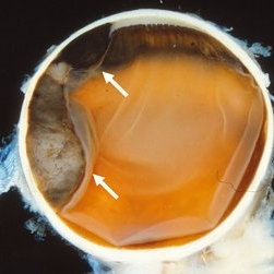
The enucleation specimen in (B) shows an amelanotic, mushroom-shaped, slightly hemorrhagic tumor near the optic nerve (arrow). The shape is due to infiltration of the retina by a rupture of the Bruch membrane. A retinal detachment artifact is present.
Condition/keywords: enucleation, mushroom-shaped
-
 Ciliary Body Melanoma
May 18 2020 by McGill University Health Centre
Uveal melanoma is the most common primary eye malignancy in adulthood, occurring mainly after age 60. The uveal tract — composed of the iris, ciliary body, and choroid — can be affected by uveal melanoma. Despite advances in treatment of the primary tumor, metastatic disease occurs in almost half of patients, generally affecting the liver and lungs via hematogenous dissemination of the primary tumor. Tumors have different levels of pigmentation, and some are amelanocytic (nonpigmented). The differential diagnosis for amelanotic choroidal melanoma is metastatic disease. Large tumors displace the lens. Of the 3 locations in the uveal tract, tumors of the ciliary body have the worst prognosis. The enucleation specimen in (A) shows a firm, dome-shaped, deeply pigmented tumor arising from the ciliary body (arrow). The lens has been removed, and a diffuse retinal detachment artifact is present.
Condition/keywords: enucleation, melanoma
-
 Ciliary Body Melanoma
May 18 2020 by McGill University Health Centre
Large tumors displace the lens. Of the 3 locations in the uveal tract, tumors of the ciliary body have the worst prognosis. This enucleation specimen shows a pigmented, bilobed, dome-shaped tumor arising from the ciliary body (arrow). The lens has been removed, and a diffuse, flat retinal detachment artifact is present.
Condition/keywords: melanoma
-
 Common Artifacts in Macroscopic Ocular Globe Evaluation
May 18 2020 by McGill University Health Centre
This sample was retrieved from a patient with a blind, painful eye. Blind, painful eye may be the end stage of several conditions including glaucoma, retinal detachment, and endophthalmitis, among others. Evisceration specimens are generally submitted in fragments. Different intraocular structures are identifiable: retina, cornea and capsular bag, choroidal tissue, and hematic material.
Condition/keywords: enucleation, evisceration, intraocular structures
-
 Ectopia Lentis
Jan 21 2021 by Jamin S. Brown, MD
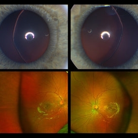
This image serial demonstrates a patient with simple ectopia lentis. Anterior segment photographs in the upper panel demonstrate nasally subluxated crystalline lenses. Widefield fundus photography shows a "pseudo-buckle" which is the result of an optical effect due to the lens subluxation (artifactual image enlargement). Also note the juvenile macular reflex in this young patient. Ectopia lentis can present isolated ("simple") or in combination with various systemic defects (Marfan's syndrome, Weil-Marchesani syndrome or Ehlers-Danlos syndrome to name a few). Isolated ectopia lentis can be hereditary and causative genes have been identified as ADAMTSL4 located on chromosome 4 and FBN1 gene located on chromosome 15. Defects in the genes cause weakness in the zonular fibers which can lead to lens dislocation. Lastly, various ocular disorders such as Aniridia, Axenfeld-Rieger, Pseudoexfoliation or Trauma may also result in lens dislocation or subluxation.
Photographer: Stefanie Palmer CRA, Retina Vitreous Surgeons of CNY
Condition/keywords: dislocated lens, ectopia lentis
-
 Enucleated Eye with Subretinal Hematoma
May 18 2020 by McGill University Health Centre
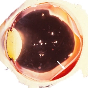
This enucleation specimen shows a subretinal hematoma (arrow). In addition, a diffuse, flat retinal detachment artifact is present. The lens is cataractous.
Condition/keywords: cataract, subretinal hemorrhage
 Loading…