 Initializing download.
-
 By McGill University Health Centre
The MUHC-McGill University
Co-author(s): Sabrina Bergeron, P. Zoroquiain, E. Esposito, S. Corredor Casas, P. Logan, A. N. Odashiro, Miguel N. Burnier, Paulina García de Alba Graue, McGill University Health Center-McGill University Ocular Pathology & Translational Research Laboratory - Uploaded on May 18, 2020.
- Last modified by Caroline Bozell on May 19, 2020.
- Rating
- Appears in
- Pigmented Tumor
- Condition/keywords
- tumor, enucleation, infiltrating ciliary body
- Description
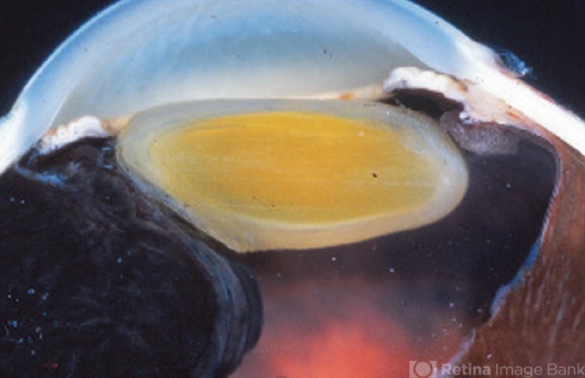
- This enucleation specimen shows a heavily pigmented, solid tumor-infiltrating the ciliary body. The tumor infiltrates the left angle, producing secondary glaucoma. Note the dislocation of the lens and the choroidal detachment artifact on the right bottom angle of the image.


---thumb.jpg/image-square;max$79,0.ImageHandler "Vitrectomy Choroidal Mass")


---thumb.jpg/image-square;max$79,0.ImageHandler "unknown")

