 Initializing download.
-
 By Gustavo Uriel Fonseca Aguirre
Hospital de la Luz - Uploaded on Apr 21, 2025.
- Last modified by Joshua Friedman on Apr 23, 2025.
- Rating
- Appears in
- Miscellaneous
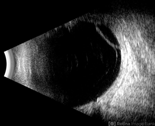
- Condition/keywords
- retinoschisis
- Photographer
- Gustavo U. Fonseca Aguirre, Hospital Conde de Valenciana, Ciudad de México
- Imaging device
- Ultrasonography device
- Description
- This B-mode longitudinal ultrasound scan reveals a peripheral temporal retinoschisis, demonstrating a characteristic thin, dome-shaped separation of the retinal layers without associated subretinal fluid or vitreous traction. The lesion shows smooth, convex contours with maintained structural integrity of both retinal layers.


")


---thumb.jpg/image-square;max$79,0.ImageHandler "Retinoschisis")


