Search results (149 results)
-
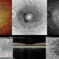
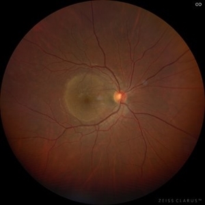
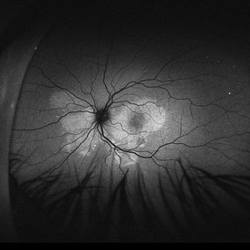
 Retinitis Pigmentosa
Oct 1 2025 by Shivankar Sen, MS, FVRS
Typical Case of Retinitis Pigmentosa - Optos Green Auto Fluorescence highlighting Peripapillary Hypoautofluorescence; a classical ring hypoautofluorescence at the macula surrounding the fovea and speckled autofluorescence in the periphery corresponding to bony spicules
Photographer: Dr. Shivankar Sen
Imaging device: Optos Daytona
Condition/keywords: fundus autofluorescence (FAF), Optos, RETINITIS PIGMENTOSA
-
 CMV Retinitis: Turning Retina into Abstract Art Since Immunosuppression
Aug 4 2025 by rohan jain
We report a case of 34 years old HIV positive male who presented with Diminution of vision in OD since 1 month .Examination of OD showed hazy media due to vitritis, diffuse yellowish-whitish retinal necrosis and retinal hemorrhages around the disc and attenuated retinal vessels.
Photographer: Dr. ROHAN JAIN
Imaging device: mirante
Condition/keywords: CMV chorioretinitis, CMV retinitis, cytomegalovirus (CMV), Cytomegalovirus Retinitis
-
 CMV Retinitis: Turning Retina into Abstract Art Since Immunosuppression
Aug 4 2025 by rohan jain
We report a case of 34 years old HIV positive male who presented with Diminution of vision in OD since 1 month. Examination of OD showed hazy media due to vitritis, diffuse yellowish-whitish retinal necrosis and retinal hemorrhages around the disc and attenuated retinal vessels.
Photographer: Dr. ROHAN JAIN
Imaging device: mirante
Condition/keywords: CMV chorioretinitis, CMV retinitis, cytomegalovirus (CMV), Cytomegalovirus Retinitis
-
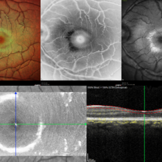
 Stargardt's Disease (Extensive)
Jul 16 2025 by Shivankar Sen, MS, FVRS
15-year-old female with complaints of defective vision for 6 years with best corrected visual acuity of 6/60; N36 in both eyes was found to have dystrophic macula with extensive spread out pigmentary bony spicules; On Confocal blue autofluorescence shows central hypo-autofluorescence and a heterogenous pisciform background with OCT showing extensive outer retinal layer disruption with genetic report confirming ABCA4 mutation and giving us definite diagnosis of Stargardt's Disease.
Photographer: Dr. N. Haindavi
Imaging device: Heidelberg Spectralis HRA+OCT
Condition/keywords: Blue autofluroscence, Stargardts Disease
-
 Combined Occlusion with TRD
Jun 30 2025 by Shivankar Sen, MS, FVRS
Posterior Pole and Ultra-wide field Fluorescein angiogram of a 79 yr. old one eyed male revealing arterial occlusion, grossly non-perfused peripheral retina with neovascularisation elsewhere and significant tractions at the posterior pole.
Photographer: Gayathri M S, Dr. Shivankar Sen MD
Imaging device: Heidelberg Spectralis HRA+OCT
Condition/keywords: arterial occlusion, Traction retinal detachment, Vein Occlusion
-
 CRAO With Cilio-retinal Sparing-MMI
Jun 25 2025 by Shivankar Sen, MS, FVRS
A 41 year old male came with complaints of Right eye blurring of vision since a day associated with watering and redness. He had no systemic illness, though gave a history of fall from bike 1 month back at the time of which he had blunt force trauma to the right side of the face. BCVA was 3/60, less than N36 in the right eye and 6/6, N6 in the left eye. Right eye had Marcus Gunn Pupil with clear lens, Left eye was within normal limits. IOP was normal; 16 in OD and 18 in OS. Retina evaluation revealed CRAO in the right eye with cilio-retinal artery sparing. Left eye was unremarkable Image Details Left to Right (Top 2 rows) Multicolor Reflectance Image (Blue-green enhanced 55 degree) revealing cilioretinal spared retinal stroma and a characteristic Cherry Red Spot; Green Reflectance showing corresopnding dark gray area with spared perfusion and black spot consistent with Cherry Red Spot on multicolor 2nd Row - 35 degree image (Multicolor Standard Reflectance and Green Reflectance) 3rd Row - SD-OCT revealing acute moderate CRAO findings with Middle retinal layer opacification and prominent middle limiting membrane (p-MLM) sign; Inner retinal layer opacification and prominent retinal pigment epithelium at the fovea with Diminished inner retinal layer stratification
Photographer: Gayathri M S
Imaging device: Heidelberg Spectralis HRA+OCT
Condition/keywords: CRAO with cilioretinal sparing, multicolor, multimodal imaging, OCT biomarkers, reflectance
-
 Berlins Edema - Multimodal Imaging
Jun 25 2025 by Shivankar Sen, MS, FVRS
A 22 year old female came with history of injury to her left eye with a badminton racquet butt cap an hour before presentation On examination, she was found to have right eye 6/6;N6 vision and within normal limits, left eye 6/9;N6 vision, cells1+ in the anterior chamber, brisk pupillary response, no vitreous reaction and sub-clinical berlin's edema at the posterior pole. Multimodal imaging revealed frank boundaries of Berlin's edema more pronounced in the nasal parafoveal region. Figure details Top (Left to Right) Multicolor Reflectance showing bright yellow ring surrounding the perifovea; Blue Reflectance (Black on white contrast) showing corresponding black ring; Green Reflectance showing a characteristic white ring (all pronounced nasally); Bottom (Left-Right) Transverse structural OCT enface image showing white ring consistent with edema OCTA inner layer segmentation from ILM to GCL Transverse corresponding OCTA revealing faint hypo ring within perifoveal capillary bed
Photographer: Gayathri M S
Imaging device: Heidelberg Spectralis HRA+OCT
Condition/keywords: blue reflectance, En Face OCTA, enface imaging, multicolor, oct, reflectance
-
 Berlins
Jun 25 2025 by Shivankar Sen, MS, FVRS
A 22 year old female came with history of injury to her left eye with a badminton racquet butt cap an hour before presentation On examination, she was found to have right eye 6/6;N6 vision and within normal limits, left eye 6/9;N6 vision, cells1+ in the anterior chamber, brisk pupillary response, no vitreous reaction and sub-clinical berlin's edema at the posterior pole. Multimodal imaging revealed frank boundaries of Berlin's edema more pronounced in the nasal parafoveal region. Figure details Top (Left to Right) Multicolor Reflectance showing bright yellow ring surrounding the perifovea; Blue Reflectance (Black on white contrast) showing corresponding black ring; Green Reflectance showing a characteristic white ring (all pronounced nasally); Bottom (Left-Right) Transverse structural OCT enface image showing white ring consistent with edema OCTA inner layer segmentation from ILM to GCL
Photographer: Gayathri M S
Imaging device: Heidelberg Spectralis HRA+OCT
Condition/keywords: blue reflectance, En Face OCTA, multicolor
-
 Cytomegalovirus Retinitis
Jun 23 2025 by César Adrián Gómez Valdivia, MD
Fundus Photograph of a 28 year-old male patient diagnosed with Cytomegalovirus Retinitis. Findings were unilateral. Visual acuity was 20/25 on SC. CMV is the most common opportunistic ocular pathogen in advanced HIV infection, typically occurring when CD4+ T-cell counts fall below 50 cells/µL
Photographer: @eyemissu2
Imaging device: California ICG OPTOS
Condition/keywords: Cytomegalovirus Retinitis
-
 CMV Retinitis
Jun 14 2025 by César Adrián Gómez Valdivia, MD
Fundus Photograph of a 28YO male patient diagnosed with Cytomegalovirus Retinitis. Findings were unilateral. Visual acuity was 20/25 on SC. CMV is the most common opportunistic ocular pathogen in advanced HIV infection, typically occurring when CD4+ T-cell counts fall below 50 cells/µL
Photographer: @eyemissu2
Imaging device: California ICG OPTOS
Condition/keywords: CMV retinitis
-
 Berlin's Edema
Jun 12 2025 by Shivankar Sen, MS, FVRS
A 22 year old male came with history of sports injury to the right eye with the nose of shuttlecock while playing badminton. On examination, right eye anterior segment shows conjunctival congestion with brisk pupillary reaction and quiet anterior chamber. His best corrected visual acuity was 6/12; N6 in the right eye and 6/6; N6 in the left eye. Retinal examination revealed OD Berlin's Edema, OS within normal limits. Image Description (From Left to Right) Multicolor Reflectance (Blue-Green Enhanced) shows well defined yellowish discoloration Green reflectance and blue reflectance show corresponding whitish discoloration at the area of edema
Photographer: Dr. Shivankar Sen
Imaging device: Heidelberg Spectralis HRA+OCT
Condition/keywords: Shuttlecock Injury
-
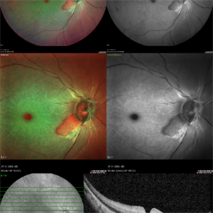
 Multi-modal Imaging of Type - 1 CNVM
May 30 2025 by Shivankar Sen, MS, FVRS
Multimodal Imaging of a case of Polypoidal Choroidal Vasculopathy Multicolor Reflectance showing multiple green-hyper-fringent lesions in the macular region (Up Left) Infra-red Autofluorescence and Blue Autofluorescence showing hypo-autofluorescent areas correspondingly revealing the exact extent of the sub-RPE Lesion (Down left and right respectively) Optical Coherence Tomography - Enhanced Depth Imaging showing Thumb-shaped Pigment Epithelial Detachment with presence of Sub-retinal fluid and Hyper-reflective foci (Top Right)
Photographer: Dr. Shivankar Sen
Imaging device: Heidelberg Spectralis HRA+OCT
Condition/keywords: Blue autofluroscence, CNVM, multicolor, near infrared autofluorescence (NIRAF), PCV, reflectance
-
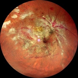
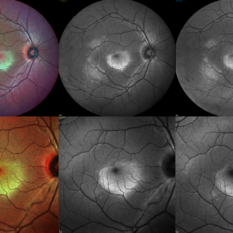
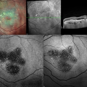
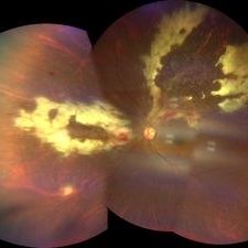
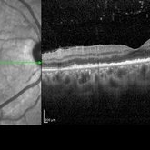 Posterior Placoid Chorioretinitis
Mar 9 2025 by Oscar Francisco Miranda, MD
A 36-year-old male with bilateral visual loss of 3 months' duration, with no relevant medical history on inquiry. A round-shaped lesion with well-defined borders and a yellowish-white color is observed in the macula of both eyes, accompanied by vitreous cellularity. The macular OCT shows a dentate RPE. The VDRL, FTA-ABS, and HIV tests were positive.
Photographer: Oscar Francisco Miranda-Gómez
Imaging device: Heidelberg Spectralis
Condition/keywords: acute syphilitic posterior placoid chorioretinitis, OCT, Ocular syphilis
-
 Posterior Placoid Chorioretinitis
Mar 9 2025 by Oscar Francisco Miranda, MD
A 36-year-old male with bilateral visual loss of 3 months' duration, with no relevant medical history on inquiry. A round-shaped lesion with well-defined borders and a yellowish-white color is observed in the macula of both eyes, accompanied by vitreous cellularity. The macular OCT shows a dentate RPE. The VDRL, FTA-ABS, and HIV tests were positive.
Photographer: Oscar Francisco Miranda-Gómez
Imaging device: Autofluorescence Zeiss Clarus 700
Condition/keywords: acute posterior placoid chorioretinitis, Autofluorescence, ocular syphilis
-
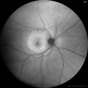
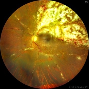
 Posterior Placoid Chorioretinitis
Mar 9 2025 by Oscar Francisco Miranda, MD
A 36-year-old male with bilateral visual loss of 3 months' duration, with no relevant medical history on inquiry. A round-shaped lesion with well-defined borders and a yellowish-white color is observed in the macula of both eyes, accompanied by vitreous cellularity. The macular OCT shows a dentate RPE. The VDRL, FTA-ABS, and HIV tests were positive.
Photographer: Oscar Francisco Miranda-Gómez
Imaging device: Zeiss Clarus 700
Condition/keywords: acute posterior placoid chorioretinitis, Ocular syphilis
-
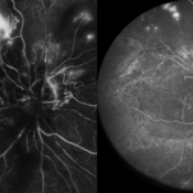
 Acute Syphilitic Posterior Placoid Chorioretinitis
Oct 20 2024 by César Adrián Gómez Valdivia, MD
Fundus autofluorescence image of an acute syphilitic posterior placoid chorioretinitis found in a HIV positive 28 YO male patient with suspected neurosyphilis. A beautiful butterfly autofluorescence pattern can be appreciated.
Photographer: @eyemissu2
Imaging device: California ICG OPTOS
Condition/keywords: acute syphilitic posterior placoid chorioretinitis
-
 Acute Syphilitic Posterior Placoid Chorioretinitis
Oct 16 2024 by César Adrián Gómez Valdivia, MD
Fundus autofluorescence image of an acute syphilitic posterior placoid chorioretinitis found in a HIV positive 28 YO male patient with suspected neurosyphilis. A beautiful butterfly autofluorescence pattern can be appreciated.
Photographer: @eyemissu2
Imaging device: California ICG OPTOS
Condition/keywords: acute syphilitic posterior placoid chorioretinitis, chorioretinitis, syphilis
-
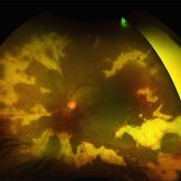
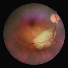
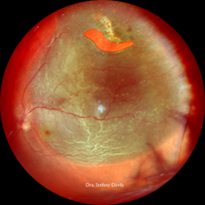 Rhegmatogenous Retinal Detachment
Aug 22 2024 by STEFANY DAVILA
Fundus photograph of a 64-year-old male patient, HIV positive for 20 years, diagnosed with rhegmatogenous retinal detachment in the upper temporal sector from M7 to M1 in the left eye with a horseshoe tear in M10.
Photographer: Stefany Dávila, Instituto Mexicano de Oftalmología, Santiago de Querétaro
Imaging device: MIRANTE NIDEK
Condition/keywords: retina, Retinal Detachment, rhegmatogenous retinal detachment
-
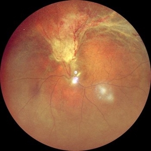 RD with PVR in CMV Retinitis in an HIV Positive Patient
Jul 31 2024 by Tejaswita Verma
Fundus photograph of a 48 year old male with CF 1.5 mt vision having history of CMV retinitis, on HAART with CD4 count 81, showing retinal detachment with proliferative vitreoretinopathy changes. He was advised pars plana vitrectomy with silicon oil infusion.
Photographer: DR. TEJASWITA VERMA
Imaging device: MIRANTE
Condition/keywords: CMV retinitis with retinal detachment, HIV
-
 Weiss Ring
Apr 22 2024 by SHIVANG CHAURASIA
Fundus photograph of a 68-year-old female with complaints of floaters.
Photographer: Dr SHIVANG CHAURASIA, GSVM MEDICAL COLLEGE, KANPUR, UTTAR PRADESH, INDIA
Imaging device: SMARTPHONE FUNDOSCOPY- IPHONE12
Condition/keywords: posterior vitreous detachment, Weiss ring
-
 Cheese Pizza Pie Appearance in CMV Retinitis
Mar 30 2024 by KANWALJEET HARJOT MADAN, M.S. (Ophthalmology); FAICO (Vitreous - Retina)
This is Fundus Photograph of left eye of 53 year male depicting an area of Retinal Necrosis with few Retinal Haemorrhages suggestive of CMV Retinitis. Areas of Perivascular Exudation also seen. On investigations, the patient was found to be HIV positive. He was started on Anti Retro Viral treatment after physician opinion.
Photographer: Dr. Kanwaljeet Harjot Madan, Thind Eye Hospital, Jalandhar City (Punjab) INDIA.
Imaging device: Zeiss Fundus Camera
Condition/keywords: AIDS, cytomegalovirus (CMV), retinitis
-
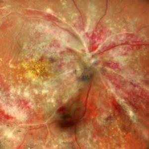
 CMV Retinitis
Feb 17 2024 by Eloy Mata-Cortes, MD
Fundus photograph of left eye showing Cytomegalovirus retinitis of a 40-year-old male with positive HIV history. He presented with CD4 cell count of 50 cells/mm3 and decreased vision of left eye. In the photograph we can see the three typical patterns in this retinitis: a hemorrhagic appearance in superior temporal arcade and between nasal arcades, granular pattern in superior temporal retina, and a “frosted branch” angiitis surrounding the retinal vessels in nasal and superior retina.
Photographer: Eloy Mata-Cortes, Instituto Mexicano de Oftalmologia, Queretaro, Mexico
Imaging device: Clarus 700
Condition/keywords: CMV retinitis, cytomegalovirus (CMV), frosted branch angiitis, Frosted Branch Angitis
-
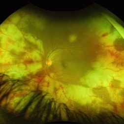
 Ocular toxoplasmosis
Mar 5 2023 by Sergio Emilio Sifuentes Renteria, MD
Color fundus photograph of the right eye of a patient with HIV-infection and concomitant ocular toxoplasmosis.
Photographer: Sergio Emilio Sifuentes Rentería - Clínica Especializada Condesa Iztapalapa
Condition/keywords: HIV, infectious uveitis, posterior uveitis, toxoplasmosis, toxoplasmosis chorioretinitis
-
 Proliferative diabetic retinopathy with fibrous proliferation over disc
Nov 4 2022 by T. P . VIGNESH, MBBS,MS
SD-OCT of a 60 year old man with proliferative diabetic retinopathy post PRP laser, revealing regressed fibrous proliferation attached to the disc .
Photographer: Shivanath
Imaging device: Heidelberg Spectralis
Condition/keywords: proliferative diabetic retinopathy (PDR)
 Loading…