 Initializing download.
-
 By Eloy Mata-Cortes, MD
Instituto Mexicano de Oftalmología
Co-author(s): Veronica Romero-Morales, Instituto Mexicano de Oftalmologia, Queretaro, Mexico - Uploaded on Feb 17, 2024.
- Last modified by Joshua Friedman on Feb 19, 2024.
- Rating
- Appears in
- Miscellaneous
- Condition/keywords
- CMV retinitis, cytomegalovirus (CMV), frosted branch angiitis, Frosted Branch Angitis
- Photographer
- Eloy Mata-Cortes, Instituto Mexicano de Oftalmologia, Queretaro, Mexico
- Imaging device
-
Fundus camera
Clarus 700 - Description
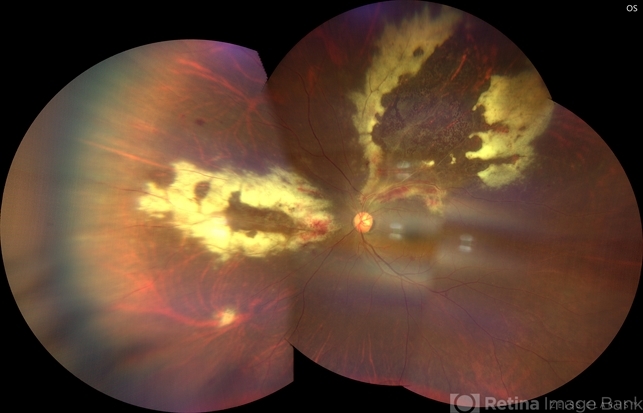
- Fundus photograph of left eye showing Cytomegalovirus retinitis of a 40-year-old male with positive HIV history. He presented with CD4 cell count of 50 cells/mm3 and decreased vision of left eye. In the photograph we can see the three typical patterns in this retinitis: a hemorrhagic appearance in superior temporal arcade and between nasal arcades, granular pattern in superior temporal retina, and a “frosted branch” angiitis surrounding the retinal vessels in nasal and superior retina.


---thumb.jpg/image-square;max$79,0.ImageHandler "CMV Frosted Branch Angitis")
---thumb.jpg/image-square;max$79,0.ImageHandler "Frosted Branch Angiitis")
---thumb.jpg/image-square;max$79,0.ImageHandler "Frosted Branch Angiitis")
---thumb.jpg/image-square;max$79,0.ImageHandler "Frosted Branch Angiitis")

---thumb.jpg/image-square;max$79,0.ImageHandler "Frosted Branch Angiitis")
---thumb.jpg/image-square;max$79,0.ImageHandler "Frosted Branch Angiitis")
---thumb.jpg/image-square;max$79,0.ImageHandler "Frosted Branch Angiitis")
---thumb.jpg/image-square;max$79,0.ImageHandler "CMV Retinitis Granular Type")
---thumb.jpg/image-square;max$79,0.ImageHandler "Possible CMV Retinitis with Frosted Branch Angiitis")