Search results (138 results)
-
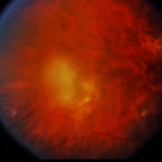
 CMV Retinitis in AIDS Patient
Dec 12 2019 by McGill University Health Centre
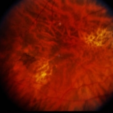
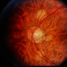
Fundus photograph of a 32-year-old man with HIV infection and 100 CD4+ cells count. Several areas of retinal necrosis interspersed with areas of hemorrhage around blood vessels can be observed.
Photographer: Miguel N. Burnier, McGill University Health Center-McGill University Ocular Pathology & Translational Research Laboratory
Imaging device: Fundoscopy
Condition/keywords: AIDS, cytomegalovirus (CMV), HIV, retinitis
-
 CMV Retinitis in AIDS Patient
Dec 12 2019 by McGill University Health Centre
Fundus photograph of a 32-year-old man with HIV infection and 100 CD4+ cells count. Several areas of retinal necrosis interspersed with areas of hemorrhage around blood vessels can be observed.
Photographer: Miguel N. Burnier, McGill University Health Center-McGill University Ocular Pathology & Translational Research Laboratory
Imaging device: Fundoscopy
Condition/keywords: AIDS, cytomegalovirus (CMV), HIV, retinitis
-
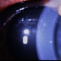
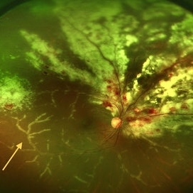 CMV Retinitis with Frosted Branch Angiitis
Sep 23 2020 by Nimesh A. Patel, MD, FASRS
Fundus photo showing peri-vascular inflammation of both arteries and veins with translucent exudation (yellow arrow). Superior nasally, there is classic retinal whitening with retinal hemorrhages superior. This patient was found to have a low CD4 count and a diagnosis of AIDS was made.
Condition/keywords: cytomegalovirus (CMV), HIV, uveitis
-
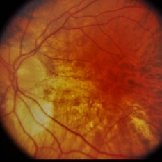
 CMV retinitis/ After treatment
Mar 13 2015 by Niloofar Piri, MD
The same patient one month after systemic treatment with Gancyclovvir and HAART ; resolved retinitis and hemorrhages, granular pattern remains as the outer retina is damaged.
Photographer: Angela Anderson
Condition/keywords: CMV retinitis, HIV
-
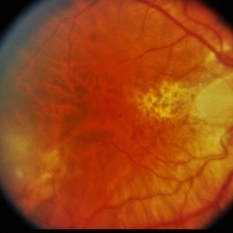
 CMV Retinitis/ Before Treatment
Mar 13 2015 by Niloofar Piri, MD
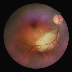
Fundus photograph of the left eye of a 40-year-old Caucasian female with history of positive HIV test for 23 years. She has been off HAART therapy for the past 2 years and presented with decreased vision OS and upper visual field defect. On examination, she had trace cells in anterior vitreous , hemorrhagic retinitis which starts around the optic nerve and extending to inferotemporal arcade with secondary inferotemporal BRVO; in temporal periphery , she had granular pattern of CMV retinitis which is a manifestation of outer retina involvement.
Photographer: Angela Anderson
Condition/keywords: CMV retinitis, HIV
-
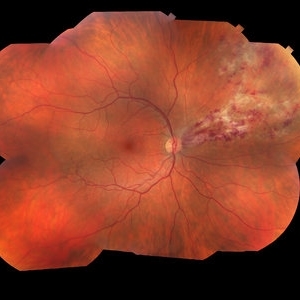 Cytomegalovirus Retinitis
Jan 16 2018 by Olivia Rainey
Color fundus montage of an 37-year-old, HIV positive male with CMV retinitis affecting his right eye. Patient's vision was sc20/20-1. He received an intravitreal Ganciclovir injection as well. The referring physcian suspects his condition is secondary to his chemotherapy for large B cell lymphoma or stomach cancer. The patient had not started taking oral Valgancyclovir.
Photographer: Olivia Rainey
Imaging device: Topcon 50dx
Condition/keywords: CMV retinitis, color fundus photograph, cytomegalovirus (CMV), HIV, montage
-
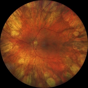 Didanosine Toxicity
Jan 27 2020 by Nimesh A. Patel, MD, FASRS
Patient with history of HIV treated with didanosine. Developed gyrate like peripheral retinal atrophy with central sparing. Vision is 20/25
Imaging device: Clarus
Condition/keywords: AIDS, didanosine, HIV
-
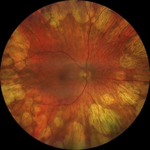 Didanosine Toxicity
Jan 27 2020 by Nimesh A. Patel, MD, FASRS
Patient with history of HIV treated with didanosine. Developed gyrate like peripheral retinal atrophy with central sparing. Vision is 20/25
Imaging device: Clarus
Condition/keywords: AIDS, didanosine, HIV
-
 Disseminated Chorioretinitis With Unknown Etiology
Apr 5 2018 by Kim Barrett
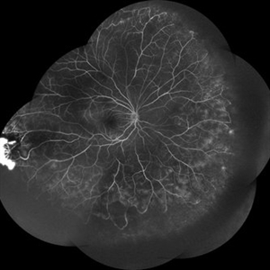
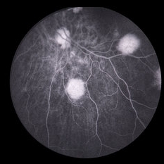
Ultra-wide field fluorescein angiogram of a 31-year-old female with intermittent pain in her left eye. Her condition has been managed in Liberia until recently when she moved to the United States. She suffers from multiple modalities including central retinal artery occlusion, posterior synechiae of the iris, interstitial keratitis, disseminated chorioretinitis, as well as HIV. An infectious cause is high on the differential in light of her HIV status. DDx: hypertensive crisis, an embolism (? IV drug use), coagulopathy, trauma, infectious. Blood work was normal. Her current vision is 20/30 right eye and 20/400 left eye.
Photographer: Kim Barrett, COA
Imaging device: Optos
Condition/keywords: central retinal artery occlusion (CRAO), chorioretinal scar, ciliary artery sparring, disseminated chorioretinitis, HIV, left eye, optic atrophy, staining
-
 HIV - Toxoplasmosis
Jan 20 2015 by David Callanan, MD
37-year-old Hispanic male, HIV - toxoplasmosis.
Condition/keywords: HIV, toxoplasmosis
-
 HIV - Toxoplasmosis
Jan 20 2015 by David Callanan, MD
37-year-old Hispanic male, HIV - toxoplasmosis.
Condition/keywords: HIV, toxoplasmosis
-
 HIV - Toxoplasmosis
Jan 20 2015 by David Callanan, MD
37-year-old Hispanic male, HIV - toxoplasmosis.
Condition/keywords: HIV, toxoplasmosis
-
 HIV - Toxoplasmosis
Jan 20 2015 by David Callanan, MD
37-year-old Hispanic male, HIV - toxoplasmosis.
Condition/keywords: HIV, toxoplasmosis
-
 HIV - Toxoplasmosis
Jan 20 2015 by David Callanan, MD
37-year-old Hispanic male, HIV - toxoplasmosis.
Condition/keywords: HIV, toxoplasmosis
-
 HIV Retinopathy with Very Early Cytomegalovirus Retinitis
Sep 27 2012 by Jeffrey G. Gross, MD, FASRS
HIV retinopathy with very early CMV retinitis, inferotemporal arcade.
Condition/keywords: HIV, inferotemporal arcade, retinopathy
-
 NVE in an HIV Positive Case
Sep 26 2021 by Nivesh Gupta
FA Montage of a 22-year-old female with Neovascularization Elsewhere
Photographer: DR. NIVESH GUPTA, RETINA FOUNDATION, AHMEDABAD
Imaging device: NIDEK MIRANTE
Condition/keywords: HIV, neovascularization elsewhere (NVE)
-
 Ocular toxoplasmosis
Mar 5 2023 by Sergio Emilio Sifuentes Renteria, MD
Color fundus photograph of the right eye of a patient with HIV-infection and concomitant ocular toxoplasmosis.
Photographer: Sergio Emilio Sifuentes Rentería - Clínica Especializada Condesa Iztapalapa
Condition/keywords: HIV, infectious uveitis, posterior uveitis, toxoplasmosis, toxoplasmosis chorioretinitis
-
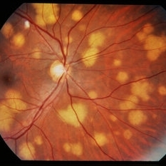
 Pneumocystis Carinii Choroiditis
Dec 12 2019 by McGill University Health Centre
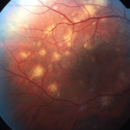
38-year-old HIV positive patient with AIDS. Fundoscopy shows large and small subretinal (choroidal) nodular lesions through out the retina in particular between the arcades.
Photographer: Miguel N. Burnier, McGill University Health Center-McGill University Ocular Pathology & Translational Research Laboratory
Imaging device: Fundoscopy
Condition/keywords: AIDS, choroidal lesions, HIV
-
 Pneumocystis Carinii Choroiditis
Dec 12 2019 by McGill University Health Centre
38-year-old HIV positive patient with AIDS. Fundoscopy showing large and small subretinal (choroidal) nodular lesions throughout the retina in particular between the arcades
Photographer: Miguel N. Burnier, McGill University Health Center-McGill University Ocular Pathology & Translational Research Laboratory
Imaging device: Fundoscopy
Condition/keywords: AIDS, choroidal lesions, HIV, pneumocystis carinii choroiditis
-
 Pneumocystis Carinii Choroiditis
Dec 18 2014 by H. Michael Lambert, MD
Pneumocystis carinii choroiditis in a patient with HIV and AIDS.
Condition/keywords: AIDS, HIV, pneumocystis carinii choroiditis
-
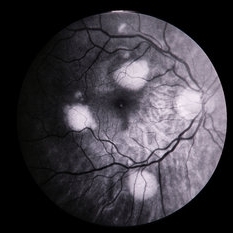
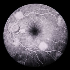
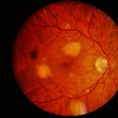 Pneumocystis Carinii Choroiditis / HIV
Feb 27 2014 by David Callanan, MD
32-year-old white male, +HIV; 20/40 OU.
Condition/keywords: choroiditis, HIV, pneumocystis
-
 Pneumocystis Carinii Choroiditis / HIV
Feb 27 2014 by David Callanan, MD
32-year-old white male, +HIV; 20/40 OU.
Condition/keywords: choroiditis, HIV, pneumocystis
-
 Pneumocystis Carinii Choroiditis / HIV
Feb 27 2014 by David Callanan, MD
32-year-old white male, +HIV; 20/40 OU.
Condition/keywords: choroiditis, HIV, pneumocystis
-
 Pneumocystis Carinii Choroiditis / HIV
Feb 27 2014 by David Callanan, MD
32-year-old white male, +HIV; 20/40 OU.
Condition/keywords: choroiditis, HIV, pneumocystis
-
 Pneumocystis Carinii Choroiditis / HIV
Feb 27 2014 by David Callanan, MD
32-year-old white male, +HIV; 20/40 OU.
Condition/keywords: choroiditis, HIV, pneumocystis
 Loading…