Search results (149 results)
-
---thumb.jpg/image-square;max$300,300.ImageHandler) HIV Retinopathy
HIV Retinopathy
Feb 27 2013 by Henry J. Kaplan, MD
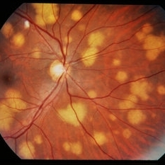
HIV retinopathy, multiple cotton wool spots, right eye. #1
Condition/keywords: cotton wool spots, HIV retinopathy
-
 HIV retinopathy with resolving CMV retinitis - left eye
Jan 11 2013 by Alex P. Hunyor, MD
HIV retinopathy and resolving CMV retinitis, left eye. 36-year-old male with HIV/AIDS. Multiple cotton wool spots due to HIV microangopathy, and an area of resolving CMV retinitis superior to the fovea (patient undergoing treatment with IV ganciclovir).
Condition/keywords: CMV retinitis, HIV retinopathy
-
 HIV retinopathy RE
Jan 11 2013 by Alex P. Hunyor, MD
HIV retinopathy, right eye. 36-year-old male with HIV/AIDS.
Condition/keywords: HIV retinopathy
-
 HIV Retinopathy with Very Early Cytomegalovirus Retinitis
Sep 27 2012 by Jeffrey G. Gross, MD, FASRS
HIV retinopathy with very early CMV retinitis, inferotemporal arcade.
Condition/keywords: HIV, inferotemporal arcade, retinopathy
-
 Pneumocystis Carinii Choroiditis
Dec 18 2014 by H. Michael Lambert, MD
Pneumocystis carinii choroiditis in a patient with HIV and AIDS.
Condition/keywords: AIDS, HIV, pneumocystis carinii choroiditis
-
---thumb.jpg/image-square;max$300,300.ImageHandler) HIV Retinopathy
HIV Retinopathy
Feb 27 2013 by Henry J. Kaplan, MD
HIV retinopathy, left eye: multiple cotton wool spots. #2
Condition/keywords: cotton wool spots, HIV retinopathy
-
 HIV Retinopathy
Aug 20 2014 by Andree Henaine-Berra, MD
Fundus photograph of the right eye of a HIV-positive male patient. The image shows multiple cotton wool spots and vascular tortuosity.
Photographer: Jorge Morales, MD. Hospital General "Dr. Manuel Gea Gonzalez". Mexico City
Condition/keywords: HIV retinopathy
-
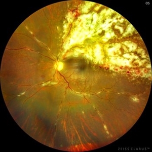
 CMV Retinitis/ Before Treatment
Mar 13 2015 by Niloofar Piri, MD
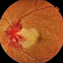
Fundus photograph of the left eye of a 40-year-old Caucasian female with history of positive HIV test for 23 years. She has been off HAART therapy for the past 2 years and presented with decreased vision OS and upper visual field defect. On examination, she had trace cells in anterior vitreous , hemorrhagic retinitis which starts around the optic nerve and extending to inferotemporal arcade with secondary inferotemporal BRVO; in temporal periphery , she had granular pattern of CMV retinitis which is a manifestation of outer retina involvement.
Photographer: Angela Anderson
Condition/keywords: CMV retinitis, HIV
-
---thumb.JPG/image-square;max$300,300.ImageHandler) Tubercular choroidal granuloma
Tubercular choroidal granuloma
Oct 26 2012 by Mallika Goyal, MD
Fundus photograph of left eye of a 43-year-old HIV infected gentleman who started antiretroviral therapy 2 months prior to visual symptoms in this eye.
Condition/keywords: tubercular choroidal granuloma
-
 Adenocarcinoma Arising from CHRPE
Sep 17 2015 by Marc C. Peden, MD
49-year-old female referred for presumed ocular melanoma. On examination was noted to have darkly pigmented lesion in the temporal retina of left eye. Lesion had characteristic scalloped edges with central lacunae, however, on ultrasonography was noted to have 1.8mm of elevation with high internal reflectivity. IVFA shows absence of dual circulation with areas of window defect. Findings were consistent with those described by Shields et al., in their April 2001 article in Archives of Ophthalmology.
Photographer: Janet Traynom
Imaging device: Optos P200MA
Condition/keywords: adenocarcinoma arising from CHRPE
-
 Tubercular choroidal granuloma
Oct 26 2012 by Mallika Goyal, MD
Fundus photograph of a 43-year-old HIV infected gentleman with choroidal granuloma improving on antiretroviral therapy, second line antitubercular therapy (for MDR TB) and steroids (for immune reconstitution syndrome)
Condition/keywords: tubercular choroidal granuloma
-
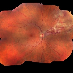 Cytomegalovirus Retinitis
Jan 16 2018 by Olivia Rainey
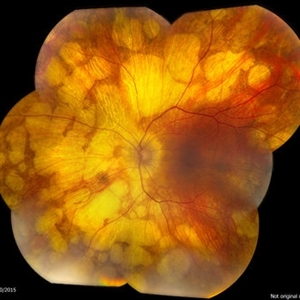
Color fundus montage of an 37-year-old, HIV positive male with CMV retinitis affecting his right eye. Patient's vision was sc20/20-1. He received an intravitreal Ganciclovir injection as well. The referring physcian suspects his condition is secondary to his chemotherapy for large B cell lymphoma or stomach cancer. The patient had not started taking oral Valgancyclovir.
Photographer: Olivia Rainey
Imaging device: Topcon 50dx
Condition/keywords: CMV retinitis, color fundus photograph, cytomegalovirus (CMV), HIV, montage
-
 Cheese Pizza Pie Appearance in CMV Retinitis
Mar 30 2024 by KANWALJEET HARJOT MADAN, M.S. (Ophthalmology); FAICO (Vitreous - Retina)
This is Fundus Photograph of left eye of 53 year male depicting an area of Retinal Necrosis with few Retinal Haemorrhages suggestive of CMV Retinitis. Areas of Perivascular Exudation also seen. On investigations, the patient was found to be HIV positive. He was started on Anti Retro Viral treatment after physician opinion.
Photographer: Dr. Kanwaljeet Harjot Madan, Thind Eye Hospital, Jalandhar City (Punjab) INDIA.
Imaging device: Zeiss Fundus Camera
Condition/keywords: AIDS, cytomegalovirus (CMV), retinitis
-
 Tubercular choroidal granuloma
Oct 26 2012 by Mallika Goyal, MD
Fundus photograph of 43-year-old HIV infected gentleman on treatment with antiretroviral therapy, second-line antitubercular medication (for MDR TB) and steroids (for immune reconstitution syndrome) with improvement in lesion characteristics.
Condition/keywords: tubercular choroidal granuloma
-
 CMV Retinitis with Frosted Branch Angiitis
Sep 23 2020 by Nimesh A. Patel, MD, FASRS
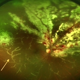
Fundus photo showing peri-vascular inflammation of both arteries and veins with translucent exudation (yellow arrow). Superior nasally, there is classic retinal whitening with retinal hemorrhages superior. This patient was found to have a low CD4 count and a diagnosis of AIDS was made.
Condition/keywords: cytomegalovirus (CMV), HIV, uveitis
-
 Disseminated Chorioretinitis With Unknown Etiology
Apr 5 2018 by Kim Barrett
Ultra-wide field fluorescein angiogram of a 31-year-old female with intermittent pain in her left eye. Her condition has been managed in Liberia until recently when she moved to the United States. She suffers from multiple modalities including central retinal artery occlusion, posterior synechiae of the iris, interstitial keratitis, disseminated chorioretinitis, as well as HIV. An infectious cause is high on the differential in light of her HIV status. DDx: hypertensive crisis, an embolism (? IV drug use), coagulopathy, trauma, infectious. Blood work was normal. Her current vision is 20/30 right eye and 20/400 left eye.
Photographer: Kim Barrett, COA
Imaging device: Optos
Condition/keywords: central retinal artery occlusion (CRAO), chorioretinal scar, ciliary artery sparring, disseminated chorioretinitis, HIV, left eye, optic atrophy, staining
-
 Syphilitic Maculopathy
Aug 27 2012 by Logan Milad Haak, MD
51 year-old HIV+ man presenting with "Gray screen over vision" RPR and FTA-Abs were positive.
Photographer: Illinois Retina Associates
Condition/keywords: acute syphilitic posterior placoid chorioretinitis
-
---thumb.JPG/image-square;max$300,300.ImageHandler) Syphilitic Maculopathy
Syphilitic Maculopathy
Aug 31 2012 by Mathew W. MacCumber, MD, PhD
51-year-old HIV+ male presented reporting "grey screen" over his left eye. RPR and FTA-Abs were positive. Patient receive IV penicillin with subsequent improvement in vision and lesion size.
Photographer: Tara Farmer
Condition/keywords: acute syphilitic posterior placoid chorioretinitis
-
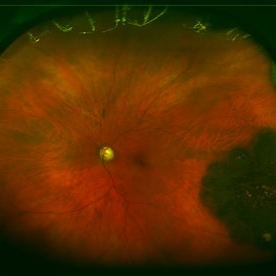
 Progressive Outer Retinal Necrosis
Nov 19 2017 by Navneet Mehrotra, DNB
42-year-old HIV positive male with reduced vision in right eye for two weeks.
Photographer: Mehul Prajapati
Imaging device: Topcon
Condition/keywords: HIV, progressive outer retinal necrosis (PORN)
-
 Syphilitic Maculopathy
Aug 31 2012 by Mathew W. MacCumber, MD, PhD
51-year-old HIV+ male presented reporting "grey screen" over his left eye. RPR and FTA-Abs were positive. Patient receive IV penicillin with subsequent improvement in vision and lesion size.
Photographer: Tara Farmer
Condition/keywords: acute syphilitic posterior placoid chorioretinitis
-
 CMV retinitis/ After treatment
Mar 13 2015 by Niloofar Piri, MD
The same patient one month after systemic treatment with Gancyclovvir and HAART ; resolved retinitis and hemorrhages, granular pattern remains as the outer retina is damaged.
Photographer: Angela Anderson
Condition/keywords: CMV retinitis, HIV
-
 Central Retinal Vein Occlusion with Cilioretinal Artery Occlusion
Oct 21 2020 by Rutul R Patel, MD Ophthalmology
Fundus photograph of left eye of 37-year-old female who presented with sudden painless loss of vision in left eye due to CRVOwith CLRAO.
Photographer: Vidhi Bavishi, Shivjyoti Eye Hospital
Imaging device: TOPCON MAESTRO
Condition/keywords: central retinal vein occlusion (CRVO), cilioretinal artery occlusion
-
 DDI Toxicity 2
Mar 5 2015 by Andrew M Hendrick, MD
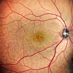
Fundus photograph of an asymptomatic 54-year-old male with a history of HIV and chronic DDI use.
Photographer: Matt Raeber, Emory University
Condition/keywords: drug toxicity
-
 Presumed Tenofovir Induced Toxicity
Nov 7 2019 by Sham Talati, DOMS
46-year-old HIV positive diabetic male with progressive bilateral decrease in vision for last 45 days. Patient had associated liver and kidney disease. Taking Tenofovir for last one year.
Photographer: Sham Talati,Retina Foundation,Ahmedabad
Imaging device: Nidek Mirante
Condition/keywords: autofluorescence imaging, drug toxicity, HIV
 Loading…