-
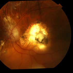
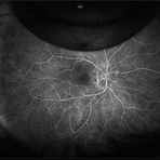
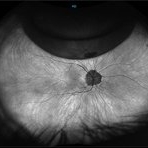
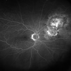
 Macula Spared Advanced Retinitis Pigmentosa 12
Apr 11 2013 by Raj K. Maturi, MD
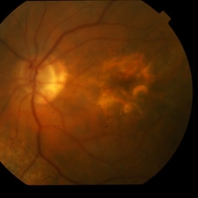
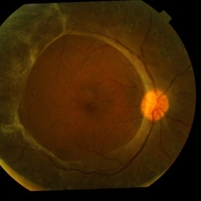
78-year-old male with advanced retinitis pigmentosa.
Photographer: Tom Steele, CRA Midwesteye Institute, Indianapolis Indiana
Condition/keywords: macula, retinitis pigmentosa
-
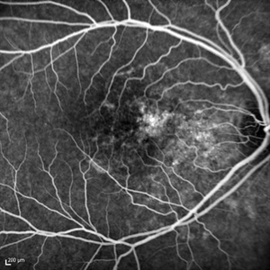
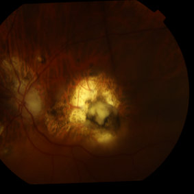
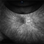
 Macula Spared Advanced Retinitis Pigmentosa 2
Apr 11 2013 by Raj K. Maturi, MD
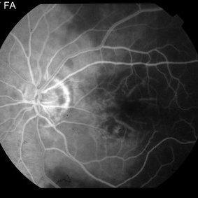
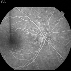
78-year-old male with advanced retinitis pigmentosa.
Photographer: Tom Steele, CRA Midwesteye Institute, Indianapolis Indiana
Imaging device: HRA
Condition/keywords: macula, retinitis pigmentosa
-
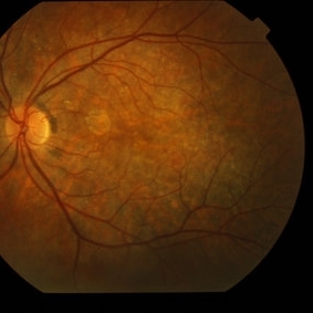
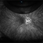
 Macula Spared Advanced Retinitis Pigmentosa 5
Apr 11 2013 by Raj K. Maturi, MD
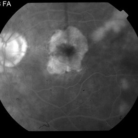
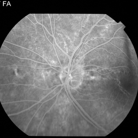
78-year-old male with advanced retinitis pigmentosa.
Photographer: Tom Steele, CRA Midwesteye Institute, Indianapolis Indiana
Imaging device: HRA
Condition/keywords: macula, retinitis pigmentosa
-
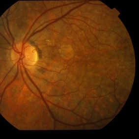
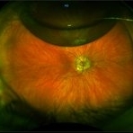
 Macula Spared Advanced Retinitis Pigmentosa 7
Apr 11 2013 by Raj K. Maturi, MD
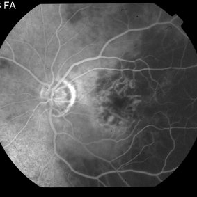
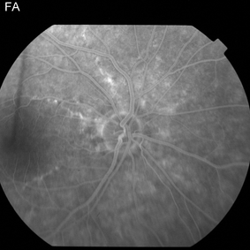
78-year-old male with advanced retinitis pigmentosa.
Photographer: Tom Steele, CRA Midwesteye Institute, Indianapolis Indiana
Condition/keywords: macula, retinitis pigmentosa
-
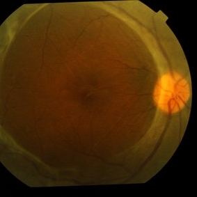
 Macula Spared Advanced Retinitis Pigmentosa 8
Apr 11 2013 by Raj K. Maturi, MD
78-year-old male with advanced retinitis pigmentosa.
Photographer: Tom Steele, CRA Midwesteye Institute, Indianapolis Indiana
Condition/keywords: macula, retinitis pigmentosa
-
 Macula Spared Advanced Retinitis Pigmentosa 11
Apr 11 2013 by Raj K. Maturi, MD
78-year-old male with advanced retinitis pigmentosa.
Photographer: Tom Steele, CRA Midwesteye Institute, Indianapolis Indiana
Condition/keywords: macula, retinitis pigmentosa
-
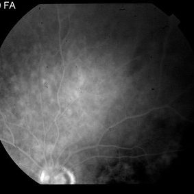
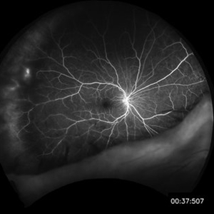
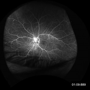
 Temporal Arteritis Delayed Choroidal Filling 014
May 15 2013 by Raj K. Maturi, MD
91 year-old female with Temporal Arteritis with delayed filling
Photographer: Tom Steele, CRA Midwest Eye Institute
-
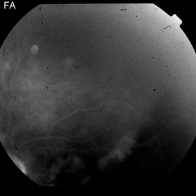
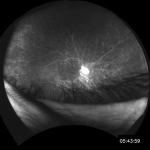
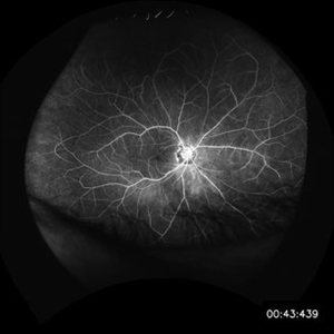
 Temporal Arteritis Delayed Choroidal Filling 004
May 15 2013 by Raj K. Maturi, MD
91-year-old female with temporal arteritis with delayed filling.
Photographer: Tom Steele, CRA Midwest Eye Institute
-
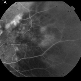
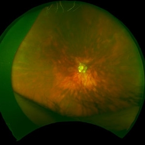
 Temporal Arteritis Delayed Choroidal Filling 005
May 15 2013 by Raj K. Maturi, MD
91-year-old female with temporal arteritis with delayed filling.
Photographer: Tom Steele, CRA Midwest Eye Institute
-
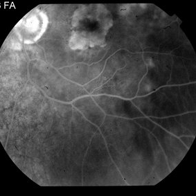
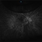
 Temporal Arteritis Delayed Choroidal Filling 006
May 15 2013 by Raj K. Maturi, MD
91-year-old female with temporal arteritis with delayed filling.
Photographer: Tom Steele, CRA Midwest Eye Institute
-
 Temporal Arteritis Delayed Choroidal Filling 008
May 15 2013 by Raj K. Maturi, MD
91-year-old female with temporal arteritis with delayed filling.
Photographer: Tom Steele, CRA Midwest Eye Institute
-
 Temporal Arteritis Delayed Choroidal Filling 009
May 15 2013 by Raj K. Maturi, MD
91-year-old female with temporal arteritis with delayed filling.
Photographer: Tom Steele, CRA Midwest Eye Institute
-
 Temporal Arteritis Delayed Choroidal Filling 010
May 15 2013 by Raj K. Maturi, MD
91-year-old female with temporal arteritis with delayed filling.
Photographer: Tom Steele, CRA Midwest Eye Institute
-
 Temporal Arteritis Delayed Choroidal Filling 011
May 15 2013 by Raj K. Maturi, MD
91-year-old female with temporal arteritis with delayed filling.
Photographer: Tom Steele, CRA Midwest Eye Institute
-
 Temporal Arteritis Delayed Choroidal Filling 012
May 15 2013 by Raj K. Maturi, MD
91-year-old female with temporal arteritis with delayed filling.
Photographer: Tom Steele, CRA Midwest Eye Institute
-
 Temporal Arteritis Delayed Choroidal Filling 013
May 15 2013 by Raj K. Maturi, MD
91-year-old female with temporal arteritis with delayed filling.
Photographer: Tom Steele, CRA Midwest Eye Institute
-
---thumb.jpg/image-square;max$300,300.ImageHandler) Stargardt 's Disease
Stargardt 's Disease
Jun 27 2013 by Raj K. Maturi, MD
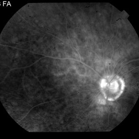
Fluorescein angiogram of a 30-year-old male with hereditary retinal dystrophy.
Photographer: Charlotte Harris COA Midwest Eye Institute Indianapolis, IN
Imaging device: Heidelberg Spectralis
Condition/keywords: Stargardt disease
-
---thumb.jpg/image-square;max$300,300.ImageHandler) Stargardt 's Disease
Stargardt 's Disease
Jun 27 2013 by Raj K. Maturi, MD
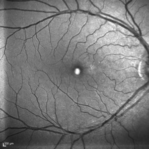
FAF of a 30-year-old male with hereditary retinal dystrophy.
Photographer: Charlotte Harris COA Midwest Eye Institute Indianapolis, IN
Imaging device: Heidelberg Spectralis
Condition/keywords: Stargardt disease
-
---thumb.jpg/image-square;max$300,300.ImageHandler) Stargardt 's Disease
Stargardt 's Disease
Jun 27 2013 by Raj K. Maturi, MD
Fluorescein angiogram of a 30-year-old male with hereditary retinal dystrophy.
Photographer: Charlotte Harris COA Midwest Eye Institute Indianapolis, IN
Imaging device: Heidelberg Spectralis
Condition/keywords: Stargardt disease
-
---thumb.jpg/image-square;max$300,300.ImageHandler) Stargardt 's Disease
Stargardt 's Disease
Jun 27 2013 by Raj K. Maturi, MD
Fluorescein angiogram of a 30-year-old male with hereditary retinal dystrophy.
Photographer: Charlotte Harris COA Midwest Eye Institute Indianapolis, IN
Imaging device: Heidelberg Spectralis
Condition/keywords: Stargardt disease
-
 Rubella Retinopathy 20
Jul 11 2013 by Raj K. Maturi, MD
48-year-old, deaf female, complains of blurry vision, mother had Rubella when she was pregnant
Photographer: Tom Steele, CRA
Imaging device: TRC 50dx
Condition/keywords: rubella retinopathy
-
 Rubella Retinopathy 1
Jul 11 2013 by Raj K. Maturi, MD
48-year-old, deaf female, complains of blurry vision, mother had Rubella when she was pregnant
Photographer: Tom Steele, CRA
Imaging device: spectralis
Condition/keywords: optical coherence tomography (OCT), rubella retinopathy, Spectralis
-
 Rubella-Retinopathy 2
Jul 11 2013 by Raj K. Maturi, MD
48-year-old, deaf female, complains of blurry vision, mother had Rubella when she was pregnant
Photographer: Tom Steele, CRA
Imaging device: spectralis
Condition/keywords: IR, rubella retinopathy, Spectralis
-
 Rubella Retinopathy 3
Jul 11 2013 by Raj K. Maturi, MD
48-year-old, deaf female, complains of blurry vision, mother had Rubella when she was pregnant.
Photographer: Tom Steele, CRA
Imaging device: spectralis
Condition/keywords: red-free, rubella retinopathy, Spectralis
-
 Rubella-Retinopathy 4
Jul 11 2013 by Raj K. Maturi, MD
48-year-old, deaf female, complains of blurry vision, mother had Rubella when she was pregnant
Photographer: Tom Steele, CRA
Imaging device: spectralis
Condition/keywords: autofluorescence imaging, rubella retinopathy, Spectralis
-
 Rubella Retinopathy 5
Jul 11 2013 by Raj K. Maturi, MD
48-year-old, deaf female, complains of blurry vision, mother had Rubella when she was pregnant.
Photographer: Tom Steele, CRA
Imaging device: spectralis
Condition/keywords: rubella retinopathy, Spectralis
-
 Rubella Retinopathy 6
Jul 11 2013 by Raj K. Maturi, MD
48-year-old, deaf female, complains of blurry vision, mother had Rubella when she was pregnant
Photographer: Tom Steele, CRA
Imaging device: spectralis
Condition/keywords: rubella retinopathy, Spectralis
-
 Rubella Retinopathy 7
Jul 11 2013 by Raj K. Maturi, MD
48-year-old, deaf female, complains of blurry vision, mother had Rubella when she was pregnant.
Photographer: Tom Steele, CRA
Imaging device: spectralis
Condition/keywords: rubella retinopathy, Spectralis
-
 Rubella Retinopathy 8
Jul 11 2013 by Raj K. Maturi, MD
48-year-old, deaf female, complains of blurry vision, mother had Rubella when she was pregnant.
Photographer: Tom Steele, CRA
Imaging device: spectralis
Condition/keywords: rubella retinopathy, Spectralis
-
 Rubella Retinopathy 9
Jul 11 2013 by Raj K. Maturi, MD
48-year-old, deaf female, complains of blurry vision, mother had Rubella when she was pregnant.
Photographer: Tom Steele, CRA
Imaging device: spectralis
Condition/keywords: optical coherence tomography (OCT), rubella retinopathy, Spectralis
-
 Rubella Retinopathy 10
Jul 11 2013 by Raj K. Maturi, MD
48-year-old, deaf female, complains of blurry vision, mother had Rubella when she was pregnant.
Photographer: Tom Steele, CRA
Imaging device: spectralis
Condition/keywords: IR, rubella retinopathy, Spectralis
-
 Rubella Retinopathy 11
Jul 11 2013 by Raj K. Maturi, MD
48-year-old, deaf female, complains of blurry vision, mother had Rubella when she was pregnant.
Photographer: Tom Steele, CRA
Imaging device: spectralis
Condition/keywords: red-free, rubella retinopathy, Spectralis
-
 Rubella Retinopathy 12
Jul 11 2013 by Raj K. Maturi, MD
48-year-old, deaf female, complains of blurry vision, mother had Rubella when she was pregnant.
Photographer: Tom Steele, CRA
Imaging device: Spectralis
Condition/keywords: autofluorescence imaging, rubella retinopathy, Spectralis
-
 Rubella Retinopathy 13
Jul 11 2013 by Raj K. Maturi, MD
48-year-old, deaf female, complains of blurry vision, mother had Rubella when she was pregnant.
Photographer: Tom Steele, CRA Midwest Eye Institute
Imaging device: Spectralis
Condition/keywords: rubella retinopathy, Spectralis
-
 Rubella Retinopathy 14
Jul 11 2013 by Raj K. Maturi, MD
48-year-old, deaf female, complains of blurry vision, mother had Rubella when she was pregnant.
Photographer: Tom Steele, CRA Midwest Eye Institute
Imaging device: Spectralis
Condition/keywords: rubella retinopathy, Spectralis
-
 Rubella Retinopathy 15
Jul 11 2013 by Raj K. Maturi, MD
48-year-old, deaf female, complains of blurry vision, mother had Rubella when she was pregnant.
Photographer: Tom Steele, CRA Midwest Eye Institute
Condition/keywords: rubella retinopathy, Spectralis
-
 Rubella Retinopathy 16
Jul 11 2013 by Raj K. Maturi, MD
48-year-old, deaf female presenting with blurry vision, mother had rubella when she was pregnant with the patient.
Photographer: Tom Steele, CRA Midwest Eye Institute Indianapolis, Indiana
Imaging device: TRC 50dx
Condition/keywords: optical coherence tomography (OCT), rubella retinopathy, Spectralis
-
 Rubella Retinopathy 17
Jul 11 2013 by Raj K. Maturi, MD
48-year-old, deaf female presenting with blurry vision, mother had rubella when she was pregnant with the patient.
Photographer: Tom Steele, CRA Midwest Eye Institute Indianapolis, Indiana
Imaging device: TRC 50dx
Condition/keywords: rubella retinopathy
-
 Rubella Retinopathy 18
Jul 11 2013 by Raj K. Maturi, MD
48-year-old, deaf female presenting with blurry vision, mother had rubella when she was pregnant with the patient.
Photographer: Tom Steele, CRA Midwest Eye Institute Indianapolis, Indiana
Imaging device: TRC 50dx
Condition/keywords: rubella retinopathy
-
 Rubella Retinopathy 19
Jul 11 2013 by Raj K. Maturi, MD
48-year-old, deaf female presenting with blurry vision, mother had rubella when she was pregnant with the patient.
Photographer: Tom Steele, CRA Midwest Eye Institute Indianapolis, Indiana
Imaging device: TRC 50dx
Condition/keywords: rubella retinopathy
-
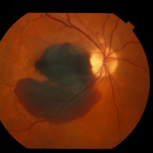
 Torpedo Maculopathy 1
Feb 26 2014 by Raj K. Maturi, MD
Color fundus of a 47-year-old female with torpedo maculopathy- atypical choroidal nevus.
Photographer: Charlotte Harris COA Midwest Eye Institute Indianapolis, Indiana
Imaging device: TOPCON 50EX
Condition/keywords: choroidal nevus, fundus photograph, torpedo maculopathy
-
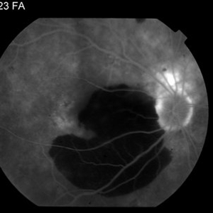
 Torpedo Maculopathy 3
Feb 26 2014 by Raj K. Maturi, MD
Fluorescein angiography of a 47-year-old female with torpedo maculopathy- atypical choroidal nevus.
Photographer: Charlotte Harris COA Midwest Eye Institute Indianapolis, Indiana
Imaging device: TOPCON 50EX
Condition/keywords: choroidal nevus, torpedo maculopathy
-
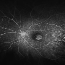
 Torpedo Maculopathy 9
Feb 26 2014 by Raj K. Maturi, MD
Fluorescein angiography of a 47-year-old female with torpedo maculopathy- atypical choroidal nevus.
Photographer: Charlotte Harris COA Midwest Eye Institute Indianapolis, Indiana
Imaging device: TOPCON 50EX
Condition/keywords: choroidal nevus, torpedo maculopathy
-
 Unilateral Retinitis Pigmentosa
May 1 2014 by Raj K. Maturi, MD
53-year-old woman with significant salt and pepper retinopathy OS.
Photographer: Tom Steele Midwesteye Institute 200 w 103rd st Indianapolis, Indiana
Condition/keywords: retinitis pigmentosa (RP) dystrophy
-
 Unilateral Retinitis Pigmentosa
May 1 2014 by Raj K. Maturi, MD
53-year-old woman with significant salt and pepper retinopathy OS.
Photographer: Tom Steele, Midwest Eye Institute
Condition/keywords: retinitis pigmentosa (RP) dystrophy
-
 Purthscher's Retinopathy
Nov 24 2014 by Raj K. Maturi, MD
Fundus images of a 21-year-old woman with Purtscher's retinopathy. Mild visual improvement 20/200 OU vs. 20/400 OU.
Photographer: Charlotte Harris Midwest Eye Institue Indianapolis, Indiana
Condition/keywords: Purtscher's retinopathy
-
 Purtscher's Retinopathy
Nov 24 2014 by Raj K. Maturi, MD
Fundus images of a 21-year-old woman with Purtscher's retinopathy. Mild visual improvement 20/200 OU vs. 20/400 OU.
Photographer: Charlotte Harris Midwest Eye Institue Indianapolis, Indiana
Condition/keywords: Purtscher's retinopathy
-
 Purtscher's Retinopathy
Nov 24 2014 by Raj K. Maturi, MD
Fundus images of a 21-year-old woman with Purtscher's retinopathy. Mild visual improvement 20/200 OU vs. 20/400 OU .
Photographer: Charlotte Harris Midwest Eye Institue Indianapolis, Indiana
Condition/keywords: Purtscher's retinopathy
-
 Purtscher's Retinopathy
Nov 24 2014 by Raj K. Maturi, MD
Fundus images of a 21-year-old woman with Purtscher's retinopathy. Mild visual improvement 20/200 OU vs. 20/400 OU.
Photographer: Charlotte Harris Midwest Eye Institue Indianapolis, Indiana
Condition/keywords: Purtscher's retinopathy
-
 Wolf Jaw
Dec 4 2015 by Raj K. Maturi, MD
45-year-old female, PDR, severe traction - wolf jaw.
Condition/keywords: proliferative diabetic retinopathy (PDR), severe traction, wolf jaw
-
 Wolf Jaw
Dec 4 2015 by Raj K. Maturi, MD
45-year-old female, PDR, severe traction - wolf jaw.
Photographer: Tom Steele, Midwest Eye Institute, Indianapolis Indiana
Imaging device: 50 & 35 degree fields, TRC 50dx
Condition/keywords: proliferative diabetic retinopathy (PDR), severe traction, wolf jaw
-
 Angioid Streaks
Dec 4 2015 by Raj K. Maturi, MD
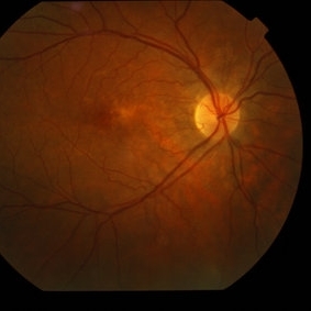
Fundus of a 24-year-old male with mild onset of blurry vision , Angioid streaks OS>OD. Observation recommended for now.
Photographer: Charlotte Harris Midwest Eye Institute Indianapolis, IN
Imaging device: TOPCON EX
Condition/keywords: angioid streaks
-
 Angioid Streaks
Dec 4 2015 by Raj K. Maturi, MD
Fundus of a 24-year-old male with mild onset of blurry vision , angioid streaks OS>OD. Observation recommended for now.
Photographer: Charlotte Harris Midwest Eye Institute Indianapolis, IN
Imaging device: TOPCON EX
Condition/keywords: angioid streaks
-
 Angioid Streaks
Dec 4 2015 by Raj K. Maturi, MD
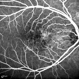
FA of a 24-year-old male with mild onset of blurry vision, angioid streaks OS>OD. Observation recommended for now.
Photographer: Charlotte Harris Midwest Eye Institute Indianapolis, IN
Imaging device: TOPCON EX
Condition/keywords: angioid streaks
-
 Angioid Streaks
Dec 4 2015 by Raj K. Maturi, MD
FA of a 24-year-old male with mild onset of blurry vision , angioid streaks OS>OD. Observation recommended for now.
Photographer: Charlotte Harris Midwest Eye Institute Indianapolis, IN
Imaging device: TOPCON EX
Condition/keywords: angioid streaks
-
 Angioid Streaks
Dec 4 2015 by Raj K. Maturi, MD
FA of a 24-year-old male with mild onset of blurry vision, angioid streaks OS>OD. Observation recommended for now.
Photographer: Charlotte Harris Midwest Eye Institute Indianapolis, IN
Imaging device: TOPCON EX
Condition/keywords: angioid streaks
-
 Angioid Streaks
Dec 4 2015 by Raj K. Maturi, MD
FA of a 24-year-old male with mild onset of blurry vision, angioid streaks OS>OD. Observation recommended for now.
Photographer: Charlotte Harris Midwest Eye Institute Indianapolis, IN
Imaging device: TOPCON EX
Condition/keywords: angioid streaks
-
 Angioid Streaks
Dec 4 2015 by Raj K. Maturi, MD
FA of a 24-year-old male with mild onset of blurry vision, angioid streaks OS>OD. Observation recommended for now.
Photographer: Charlotte Harris Midwest Eye Institute Indianapolis, IN
Imaging device: TOPCON EX
Condition/keywords: angioid streaks
-
 Angioid Streaks
Dec 4 2015 by Raj K. Maturi, MD
FA of a 24-year-old male with mild onset of blurry vision, angioid streaks OS>OD. Observation recommended for now.
Photographer: Charlotte Harris Midwest Eye Institute Indianapolis, IN
Imaging device: TOPCON EX
Condition/keywords: angioid streaks
-
 Angioid Streaks
Dec 4 2015 by Raj K. Maturi, MD
FA of a 24-year-old male presents with mild onset of blurry vision, angioid streaks OS>OD. Observation recommended for now.
Photographer: Charlotte Harris Midwest Eye Institute Indianapolis, IN
Imaging device: TOPCON EX
Condition/keywords: angioid streaks
-
 Tuberculosis at Age 12
Dec 15 2015 by Raj K. Maturi, MD
Fundus Image of a 81-year-old male - old tuberculosis scar.
Photographer: Tom Steele, CRA Midwest Eye Institute Indianapolis, Indiana
Condition/keywords: fundus photograph, macular scar, tuberculosis
-
 Tuberculosis at Age 12
Dec 15 2015 by Raj K. Maturi, MD
Fundus Image of a 81-year-old male - old tuberculosis scar.
Photographer: Tom Steele, CRA Midwest Eye Institute Indianapolis, Indiana
Condition/keywords: fundus photograph, macular scar, tuberculosis
-
 Tuberculosis at Age 12
Dec 15 2015 by Raj K. Maturi, MD
0:24 second FA of 81-year-old male - old tuberculosis scar.
Photographer: Tom Steele, CRA Midwest Eye Institute Indianapolis, Indiana
Condition/keywords: macular scar, tuberculosis
-
 Tuberculosis at Age 12
Dec 15 2015 by Raj K. Maturi, MD
0:26 second FA of 81-year-old male - old tuberculosis scar.
Photographer: Tom Steele, CRA Midwest Eye Institute Indianapolis, Indiana
Condition/keywords: macular scar, tuberculosis
-
 Tuberculosis at Age 12
Dec 15 2015 by Raj K. Maturi, MD
0:28 second FA of 81-year-old male - old tuberculosis scar.
Photographer: Tom Steele, CRA Midwest Eye Institute Indianapolis, Indiana
Condition/keywords: macular scar, tuberculosis
-
 Tuberculosis at Age 12
Dec 15 2015 by Raj K. Maturi, MD
0:31 second FA of 81-year-old male - old tuberculosis scar.
Photographer: Tom Steele, CRA Midwest Eye Institute Indianapolis, Indiana
Condition/keywords: macular scar, tuberculosis
-
 Tuberculosis at Age 12
Dec 15 2015 by Raj K. Maturi, MD
0:46 second FA of 81-year-old male old tuberclosis sca,r
Photographer: Tom Steele, CRA Midwest Eye Institute Indianapolis, Indiana
Condition/keywords: macular scar, tuberculosis
-
 Tuberculosis at Age 12
Dec 15 2015 by Raj K. Maturi, MD
2:41 minute FA of 81-year-old male - old tuberclosis scar.
Photographer: Tom Steele, CRA Midwest Eye Institute Indianapolis, Indiana
Condition/keywords: macular scar, tuberculosis
-
 Tuberculosis at Age 12
Dec 15 2015 by Raj K. Maturi, MD
4:40 minute FA of 81-year-old male - old tuberculosis scar.
Photographer: Tom Steele, CRA Midwest Eye Institute Indianapolis, Indiana
Condition/keywords: macular scar, tuberculosis
-
 Telangiectasia 1
Jan 12 2016 by Raj K. Maturi, MD
62-year-old female.
Photographer: Tom Steele, CRA Midwest Eye Institute
Condition/keywords: color photo, Optos
-
 Telangiectasia 2
Jan 12 2016 by Raj K. Maturi, MD
62-year-old female.
Photographer: Tom Steele, CRA Midwest Eye Institute
Condition/keywords: Optos
-
 Telangiectasia 3
Jan 12 2016 by Raj K. Maturi, MD
62-year-old female.
Photographer: Tom Steele, CRA Midwest Eye Institute
Condition/keywords: Optos
-
 Telangiectasia 4
Jan 12 2016 by Raj K. Maturi, MD
62-year-old female.
Photographer: Tom Steele, CRA Midwest Eye Institute
Condition/keywords: Optos
-
 Telangiectasia 5
Jan 12 2016 by Raj K. Maturi, MD
62-year-old female.
Photographer: Tom Steele, CRA Midwest Eye Institute
Imaging device: Optos
Condition/keywords: Optos
-
 Birdshot Retinopathy 1
Jan 15 2016 by Raj K. Maturi, MD
FA image of an 64-year-old white male.
Photographer: Tom Steele, CRA Midwest Eye Institute Indianapolis, Indiana
Imaging device: Optos
Condition/keywords: birdshot
-
 Birdshot Retinopathy 2
Jan 15 2016 by Raj K. Maturi, MD
FA image of an 64-year-old white male.
Photographer: Tom Steele, CRA Midwest Eye Institute Indianapolis, Indiana
Imaging device: Optos
Condition/keywords: birdshot
-
 Birdshot Retinopathy 3
Jan 15 2016 by Raj K. Maturi, MD
FA image of an 64-year-old white male.
Photographer: Tom Steele, CRA Midwest Eye Institute Indianapolis, Indiana
Imaging device: Optos
Condition/keywords: birdshot
-
 Birdshot Retinopathy 4
Jan 15 2016 by Raj K. Maturi, MD
FA image of an 64-year-old white male.
Photographer: Tom Steele, CRA Midwest Eye Institute Indianapolis, Indiana
Imaging device: Optos
Condition/keywords: birdshot chorioretinopathy
-
 Birdshot Retinopathy 5
Jan 15 2016 by Raj K. Maturi, MD
Fundus image of an 64-year-old white male.
Photographer: Tom Steele, CRA Midwest Eye Institute Indianapolis, Indiana
Imaging device: Optos
Condition/keywords: birdshot, fundus photograph
-
 Birdshot Retinopathy 6
Jan 15 2016 by Raj K. Maturi, MD
Fundus image of an 64-year-old white male.
Photographer: Tom Steele, CRA Midwest Eye Institute Indianapolis, Indiana
Imaging device: Optos
Condition/keywords: birdshot retinochoroidopathy, color fundus photograph
-
 Small Oil Bubble in Optic Nerve
May 4 2016 by Raj K. Maturi, MD
73-year-old female with persistent edema OS. Secondary to moderate neovascular AMD.
Photographer: Charlotte Harris
Imaging device: Topcon EX
Condition/keywords: optic nerve
-
 Small Oil Bubble in Optic Nerve
May 4 2016 by Raj K. Maturi, MD
73-year-old female with persistent edema OS. Secondary to moderate neovascular AMD.
Photographer: Charlotte Harris
Imaging device: Topcon EX
Condition/keywords: optic nerve
-
 Small Oil Bubble in Optic Nerve
May 4 2016 by Raj K. Maturi, MD
73-year-old female with persistent edema OS. Secondary to moderate neovascular AMD.
Photographer: Charlotte Harris
Imaging device: Topcon EX
Condition/keywords: optic nerve
-
 Small Oil Bublle in Optic Nerve
May 4 2016 by Raj K. Maturi, MD
73-year-old female with persistent edema OS. Secondary to moderate neovascular AMD.
Photographer: Charlotte Harris
Imaging device: Topcon EX
Condition/keywords: optic nerve
-
 Gas Bubble
May 21 2021 by Raj K. Maturi, MD
S/P PPV 73-year-old woman with small retinal detachment. Originally presented with decrease vision and residual vitreous floaters. Upon examination small localized retinal detachment with multiple breaks.
Photographer: Charlotte Harris ,Midwest Eye Institute 10300 N Illinois St, Carmel Indiana 46290
Imaging device: Nikon Optos California P200TDx
Condition/keywords: gas bubble
-
 Gas Bubble
May 21 2021 by Raj K. Maturi, MD
S/P PPV 73-year-old woman with small retinal detachment. Originally presented with decrease vision and residual vitreous floaters. Upon examination small localized retinal detachment with multiple breaks.
Photographer: Charlotte Harris ,Midwest Eye Institute 10300 N Illinois St, Carmel Indiana 46290
Imaging device: Nikon Optos California P200TDx
Condition/keywords: gas bubble
-
 Gas Bubble
May 21 2021 by Raj K. Maturi, MD
S/P PPV 73-year-old woman with small retinal detachment. Originally presented with decrease vision and residual vitreous floaters. Upon examination small localized retinal detachment with multiple breaks.
Photographer: Charlotte Harris ,Midwest Eye Institute 10300 N Illinois St, Carmel Indiana 46290
Imaging device: Nikon Optos California P200TDx
-
 Familial Exudative Vitreoretinopathy
Aug 30 2012 by Raj K. Maturi, MD
5-year-old male
Photographer: Tom Steele, CRA, Midwest Eye Institute
Imaging device: Topcon DX
Condition/keywords: familial exudative vitreoretinopathy (FEVR)
-
 Optic Nerve Pit
Aug 30 2012 by Raj K. Maturi, MD
Photographer: Tom Steele, CRA, Midwest Eye Institute
Imaging device: Topcon Ex
Condition/keywords: optic nerve pit
-
 Optic Nerve Pit
Aug 30 2012 by Raj K. Maturi, MD
congenital optic nerve pit with chronic pigment changes in macula due to detachment
Photographer: Tom Steele, CRA, Midwest Eye Institute
Imaging device: Topcon Ex
Condition/keywords: optic nerve pit
-
 Optic Nerve Pit
Aug 30 2012 by Raj K. Maturi, MD
Photographer: Tom Steele, CRA, Midwest Eye Institute
Imaging device: Topcon Ex
Condition/keywords: optic nerve pit
-
 Optic Nerve Pit
Aug 30 2012 by Raj K. Maturi, MD
Photographer: Tom Steele, CRA, Midwest Eye Institute
Imaging device: Topcon Ex
Condition/keywords: optic nerve pit
-
 Optic Nerve Pit
Aug 30 2012 by Raj K. Maturi, MD
Photographer: Tom Steele, CRA, Midwest Eye Institute
Imaging device: Topcon Ex
Condition/keywords: optic nerve pit
-
 Subretinal Hemorrhage
Sep 7 2012 by Raj K. Maturi, MD
Photographer: Char Harris, Midwest Eye Institute
Imaging device: TRC 50ex
-
 Subretinal Hemorrhage
Sep 7 2012 by Raj K. Maturi, MD
Photographer: Char Harris, Midwest Eye Institute
Imaging device: TRC 50ex
-
 Subretinal Hemorrhage
Sep 7 2012 by Raj K. Maturi, MD
Photographer: Char Harris, Midwest Eye Institute
Imaging device: TRC 50ex
Condition/keywords: subretinal hemorrhage
-
 Subretinal Hemorrhage
Sep 7 2012 by Raj K. Maturi, MD
Photographer: Char Harris, Midwest Eye Institute
Imaging device: TRC 50ex
Condition/keywords: subretinal hemorrhage
-
 Macular Serpiginous Choroidopathy
Sep 27 2012 by Raj K. Maturi, MD
10/21/2009
Photographer: Tom Steele, CRA
Imaging device: TRC 50ex
Condition/keywords: macula serpiginous choroidopathy
-
 Macular Serpiginous Choroidopathy
Sep 27 2012 by Raj K. Maturi, MD
10/21/2009
Photographer: Tom Steele, CRA
Imaging device: TRC 50ex
Condition/keywords: macula serpiginous choroidopathy
-
 Macular Serpiginous Choroidopathy
Sep 27 2012 by Raj K. Maturi, MD
10/21/2009
Photographer: Tom Steele, CRA
Imaging device: TRC 50ex
Condition/keywords: macula serpiginous choroidopathy
-
 Macular Serpiginous Choroidopathy
-
 Macula Serpiginous Choroidopathy
Sep 27 2012 by Raj K. Maturi, MD
9/11/2012
Photographer: Char Harris
Imaging device: HRA
Condition/keywords: IR
-
 Macular Serpiginous Choroidopathy
Sep 27 2012 by Raj K. Maturi, MD
9/11/2012
Photographer: Char Harris
Imaging device: HRA
Condition/keywords: serpiginous choroiditis
-
 Macular Serpiginous Choroidopathy
Sep 27 2012 by Raj K. Maturi, MD
9/11/2012
Photographer: Char Harris
Imaging device: HRA
Condition/keywords: red-free
-
 Macular Serpiginous Choroidopathy
-
 Macular Serpiginous Choroidopathy
Sep 27 2012 by Raj K. Maturi, MD
9/11/2012
Photographer: Char Harris
Imaging device: HRA
Condition/keywords: macula serpiginous choroidopathy
-
 Sickle Cell Retinopathy
Sep 28 2012 by Raj K. Maturi, MD
Photographer: Tom Steele, CRA
Imaging device: Optos
Condition/keywords: sickle cell retinopathy
-
 Sickle Cell Retinopathy
Sep 28 2012 by Raj K. Maturi, MD
Sickle Cell retinopathy
Photographer: Tom Steele, CRA
Imaging device: Optos
Condition/keywords: sickle cell retinopathy
-
 Coats FA
-
 Retinal Pigment Epithelium
Oct 4 2012 by Raj K. Maturi, MD
Condition/keywords: retinal pigment epithelium
-
 Retinal Pigment Epithelium
Oct 4 2012 by Raj K. Maturi, MD
Photographer: Tom Steele, CRA
Condition/keywords: optical coherence tomography (OCT), retinal pigment epithelium
-
 Retinal Pigment Epithelium
Oct 4 2012 by Raj K. Maturi, MD
Condition/keywords: retinal pigment epithelium
-
 Retinal Pigment Epithelium
Oct 4 2012 by Raj K. Maturi, MD
Photographer: Tom Steele, CRA
Condition/keywords: optical coherence tomography (OCT), retinal pigment epithelium
-
 RPE Hamartoma
Oct 18 2012 by Raj K. Maturi, MD
Photographer: Tom Steele, CRA
Imaging device: Topcon 50dx
Condition/keywords: hamartoma, red-free, retinal pigment epithelium
-
 RPE Hamartoma
Oct 18 2012 by Raj K. Maturi, MD
Photographer: Tom Steele, CRA
Imaging device: Topcon 50dx
Condition/keywords: hamartoma, retinal pigment epithelium
-
RPE Hamartoma
Oct 18 2012 by Raj K. Maturi, MD
Photographer: Stephanie Morrow
Imaging device: HRA
Condition/keywords: hamartoma, red-free, retinal pigment epithelium
-
 RPE Hamartoma
Oct 18 2012 by Raj K. Maturi, MD
Photographer: Stephanie Morrow
Imaging device: HRA
Condition/keywords: hamartoma, retinal pigment epithelium
-
 RPE Hamartoma
Oct 18 2012 by Raj K. Maturi, MD
Photographer: Stephanie Morrow
Imaging device: HRA
Condition/keywords: hamartoma, retinal pigment epithelium
-
 Stargardt's Disease
Oct 18 2012 by Raj K. Maturi, MD
Photographer: Stephanie Morrow
Imaging device: HRA
Condition/keywords: Stargardt disease
-
 Stargardt's Disease
Oct 18 2012 by Raj K. Maturi, MD
Photographer: Stephanie Morrow
Imaging device: HRA
Condition/keywords: Stargardt disease
-
 Stargardt's Disease
Oct 18 2012 by Raj K. Maturi, MD
Photographer: Stephanie Morrow
Imaging device: HRA
Condition/keywords: red-free, Stargardt disease
-
 CRVO with Choroidal Profusion Delay
Oct 18 2012 by Raj K. Maturi, MD
Photographer: Tom Steele, CRA
Imaging device: Topcon 50dx
Condition/keywords: choroidal profusion delay, FA late phase
-
 CRVO with Choroidal Profusion Delay
Oct 18 2012 by Raj K. Maturi, MD
Photographer: Tom Steele, CRA
Imaging device: Topcon 50dx
Condition/keywords: choroidal profusion delay
-
 Cat Scratch Disease
Oct 18 2012 by Raj K. Maturi, MD
Photographer: Tom Steele, CRA
Imaging device: Topcon 50dx
Condition/keywords: Bartonella bacteria, cat scratch retinitis
-
 Cat Scratch Disease
Oct 18 2012 by Raj K. Maturi, MD
Photographer: Tom Steele, CRA
Imaging device: Topcon 50dx
Condition/keywords: Bartonella bacteria, cat scratch retinitis
-
 Cat Scratch Disease
Oct 18 2012 by Raj K. Maturi, MD
Photographer: Tom Steele, CRA
Imaging device: Topcon 50dx
Condition/keywords: Bartonella bacteria, cat scratch retinitis, red-free
-
 Cat Scratch Disease
Oct 18 2012 by Raj K. Maturi, MD
Photographer: Tom Steele, CRA
Imaging device: Topcon 50dx
Condition/keywords: Bartonella bacteria, cat scratch retinitis
-
 Cat Scratch Disease
Oct 18 2012 by Raj K. Maturi, MD
Photographer: Tom Steele, CRA
Imaging device: Topcon 50dx
Condition/keywords: Bartonella bacteria, cat scratch retinitis
-
 Diabetic Retinopathy
Oct 18 2012 by Raj K. Maturi, MD
Photographer: Tom Steele, CRA
Imaging device: Optos
Condition/keywords: pan-retinal photocoagulation (PRP)
-
 Diabetic Retinopathy
Oct 18 2012 by Raj K. Maturi, MD
Photographer: Tom Steele, CRA
Imaging device: Optos
Condition/keywords: pan-retinal photocoagulation (PRP)
-
 Diabetic Retinopathy
Oct 18 2012 by Raj K. Maturi, MD
Photographer: Tom Steele, CRA
Imaging device: Optos
Condition/keywords: pan-retinal photocoagulation (PRP)
-
 Toxoplasmosis
Oct 18 2012 by Raj K. Maturi, MD
Photographer: Tom Steele, CRA
Imaging device: Optos
Condition/keywords: Optos, toxoplasmosis
-
 Toxoplasmosis
Oct 18 2012 by Raj K. Maturi, MD
Photographer: Tom Steele, CRA
Imaging device: TRC 50ex
Condition/keywords: Optos, toxoplasmosis
-
 Toxoplasmosis
Oct 18 2012 by Raj K. Maturi, MD
Photographer: Tom Steele, CRA
Imaging device: Topcon 50ex
Condition/keywords: toxoplasmosis
-
 Toxoplasmosis
Oct 18 2012 by Raj K. Maturi, MD
Photographer: Tom Steele, CRA
Imaging device: Optos
Condition/keywords: Optos, toxoplasmosis
-
 Choroidal Nevus[001]
Dec 31 2012 by Raj K. Maturi, MD
Photographer: Tom Steele CRA Midwest Eye Institute Indianapolis, In
Imaging device: Optos
Condition/keywords: choroidal nevus
-
 Choroidal Nevus[002]
Dec 31 2012 by Raj K. Maturi, MD
Photographer: Tom Steele CRA Midwest Eye Institute Indianpolis, In
Imaging device: Optos
Condition/keywords: choroidal nevus
-
 Choroidal Nevus[003]
Dec 31 2012 by Raj K. Maturi, MD
Photographer: Tom Steele CRA Midwest Eye Institute Indianpolis, In
Imaging device: Optos
Condition/keywords: choroidal nevus
-
 Choroidal Nevus[004]
Dec 31 2012 by Raj K. Maturi, MD
Photographer: Tom, Steele CRA Midwest Eye Institute Indianapolis, In
Imaging device: Optos
Condition/keywords: Optos
-
 Coats Disease
Feb 7 2013 by Raj K. Maturi, MD
8-year-old male, flourescein angiogram, late phrase 4:22 minutes, OD.
Photographer: Stephan Morrow, Midwest Eye Institute Indianapolis Indiana
Imaging device: Heidelberg Spectralis
Condition/keywords: Spectralis
-
 Coats Disease
Feb 7 2013 by Raj K. Maturi, MD
8-year-old male, Heidelberg Spectralis OCT, OD.
Photographer: Stephan Morrow, Midwest Eye Institute Indianapolis Indiana
Imaging device: Heidelberg Spectralis
Condition/keywords: optical coherence tomography (OCT), Spectralis
-
 Coats Disease
Feb 7 2013 by Raj K. Maturi, MD
8-year-old male, flourescein angiogram, venous phrase, OD.
Photographer: Stephan Morrow, Midwest Eye Institute Indianapolis Indiana
Imaging device: Heidelberg Spectralis
Condition/keywords: Spectralis
-
 Neuroretinitis
Feb 20 2013 by Raj K. Maturi, MD
28 year-old Caucasian male, presented with blurry vision OD for over 3 months.
Photographer: Tom Steele, CRA Midwest Eye Institute, Indianapolis Indiana
Imaging device: Heidelberg Spectralis
Condition/keywords: green filter, high watermark, neuroretinitis, red-free
-
 Neuroretinitis
Feb 20 2013 by Raj K. Maturi, MD
28 year-old Caucasian male, presented with blurry vision OD for over 3 months.
Photographer: Tom Steele, CRA Midwest Eye Institute, Indianapolis Indiana
Imaging device: Heidelberg Spectralis
Condition/keywords: high watermark, neuroretinitis
-
 Neuroretinitis
Feb 20 2013 by Raj K. Maturi, MD
28 year-old Caucasian male, presented with blurry vision OD for over 3 months.
Photographer: Tom Steele, CRA Midwest Eye Institute, Indianapolis Indiana
Imaging device: Heidelberg Spectralis
Condition/keywords: high watermark, neuroretinitis
-
 Neuroretinitis
Feb 20 2013 by Raj K. Maturi, MD
28 year-old Caucasian male, presented with blurry vision OD for over 3 months.
Photographer: Tom Steele, CRA Midwest Eye Institute, Indianapolis Indiana
Imaging device: Heidelberg Spectralis
Condition/keywords: IR, neuroretinitis
-
 Neuroretinitis
Feb 20 2013 by Raj K. Maturi, MD
28 year-old Caucasian male, presented with blurry vision OD for over 3 months.
Photographer: Tom Steele, CRA Midwest Eye Institute, Indianapolis Indiana
Imaging device: Heidelberg Spectralis
Condition/keywords: high watermark
A project from the American Society of Retina Specialists