 Initializing download.
-
 By J. Sebag, MD, FACS, FRCOphth, FARVO
VMR Institute for Vitreous Macula Retina - Uploaded on Sep 1, 2020.
- Last modified by Caroline Bozell on Sep 1, 2020.
- Rating
- Appears in
- Vitreous
- Condition/keywords
- embryonic eye
- Description
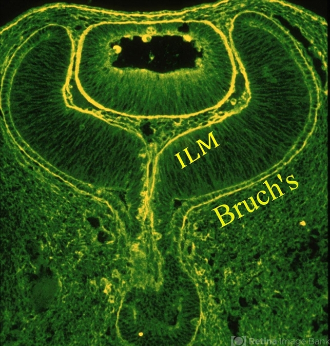
- Immunohistochemistry of a human fetal eye after invagination of the optic vesicle demonstrates basement membrane staining with fluorescein conjugated ABA lectin staining. Continuity of the membranes destined to be Bruch’s membrane and the Inner Limiting Membrane (ILM) of the retina is evident (see loops at upper aspects of this image), suggesting a common embryologic origin and similar composition. Thus, there may be important similarities during pathologies (especially neovascularization) at these two interfaces, which may provide insights into the process of pathologic cell migration and proliferation. Anterior to the ILM and behind the lens is the vascular primary vitreous. [Cover Photo - Sebag J and Hageman G: Interfaces. Farina Publishers, Rome, 2000; reprinted in Sebag J: Vitreous – in Health & Disease (J. Sebag, ed.) Springer, New York, 2014; image © Springer Nature, reprinted with permission]


