 Initializing download.
-
 By J. Sebag, MD, FACS, FRCOphth, FARVO
VMR Institute for Vitreous Macula Retina - Uploaded on Sep 1, 2020.
- Last modified by Caroline Bozell on Sep 1, 2020.
- Rating
- Appears in
- Vitreous
- Condition/keywords
- vitreous
- Description
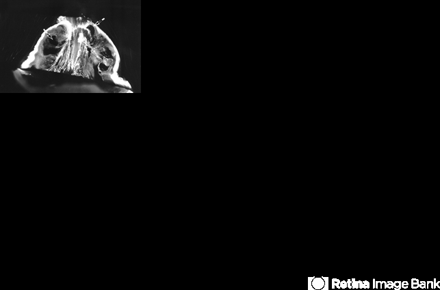
- Dark-field slit microscopy was performed on a fresh, unfixed, post-mortem human eye from an 88 year-old subject. The eye had dissection to peel off the sclera, choroid, and retina. The vitreous body remains attached to the anterior segment which is seen below, while the posterior pole is above in this image. Significant degenerative fibrous liquefaction is evident. Lacunae can be seen (arrows) that do not scatter light because they are primarily filled with liquid vitreous that is mostly hyaluronan and water, with little collagen to scatter light. Drug injection into a lacuna would likely have very different distribution than injection into an area of gel vitreous. [From Sebag J: The Vitreous - Structure, Function, and Pathobiology. Springer-Verlag, New York, 1989 (image © Springer Nature, reprinted with permission); reprinted in Sebag J: Vitreous in AMD therapy – the medium is the message (Guest Editorial). Retina 2015;35(9):1715-18]


