 Initializing download.
-
 By J. Sebag, MD, FACS, FRCOphth, FARVO
VMR Institute for Vitreous Macula Retina - Uploaded on Sep 1, 2020.
- Last modified by Caroline Bozell on Sep 1, 2020.
- Rating
- Appears in
- Vitreous
- Condition/keywords
- vitreous
- Description
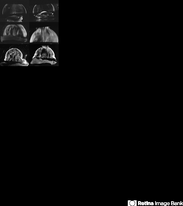
- Dark-field slit microscopy was performed on fresh, unfixed, post-mortem human eyes that had undergone dissection to peel off the sclera, choroid, and retina. The vitreous body remains attached to the anterior segment which is seen below, while the posterior pole is above in these images. The top panel demonstrates the absence of internal vitreous structures that scatter light in youth (left image from an 11 year-old girl, right image from a 14 year-old boy. The middle panel demonstrates light scattering from linear, fibrous structures that have an antero-posterior orientation with insertions into the vitreous base peripherally and the posterior vitreous cortex, typical in middle age (left image from a 56 year-old and right image from a 59 year-old). The bottom panel illustrates advance fibrous liquefaction in old age (88-year-old subject). [From Sebag J, Niemeyer M, Koss M: Anomalous PVD and vitreoschisis. In: Vitreous – in Health & Disease (J. Sebag, ed.) Springer, New York, 2014, pg. 245; image © Springer Nature, reprinted with permission]


