-
 Lamellar Structure of the Primate Posterior Vitreous Complex
Sep 3 2020 by J. Sebag, MD, FACS, FRCOphth, FARVO
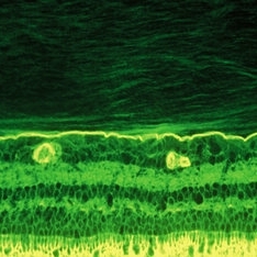
Immunohistochemistry of a monkey eye imaged with fluorescein conjugated ABA lectin staining demonstrates the lamellar structure pf the posterior vitreous cortex. During anomalous PVD, there can be splitting between these lamellae, a phenomenon known as vitreoschisis. (original magnification = 400x)
Condition/keywords: vitreous
-
 Vitreoschisis
Sep 3 2020 by J. Sebag, MD, FACS, FRCOphth, FARVO
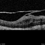
OCT of the left eye in a patient with macular pucker (see SLO image below to right) demonstrates splitting of the posterior vitreous cortex in two separate places. Tangential traction caused thickening of the underlying macula. [For histopathology see: Gupta P, Yee KMP, Garcia P, Rosen RB, Parikh J, Hageman GS, Sadun AA, Sebag J: Vitreoschisis in macular diseases. Brit J Ophthalmol 2011;95(3):376-80]
Condition/keywords: vitreoschisis
-
 Vascular Primary Vitreous
Sep 8 2020 by J. Sebag, MD, FACS, FRCOphth, FARVO
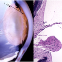
The hyaloid artery (“3”, left) feeds the vasa hyaloidea propria (“2”, left) which anastomosis with the tunica vasculosa lentis (“1”, left). The histologic section to the right is stained with H & E (bar = 100 uM) [Left: from Yee at al.: Vitreous cytokines and regression of the fetal hyaloid vasculature. In: Vitreous – in Health & Disease. Springer, New York, 2014; pg. 42 (image © Springer Nature, reprinted with permission) Right: from Sebag J: Vitreous and vitreo-retinal interface. In: Ryan’s Retina 6th edition (A. Schachat, ed.) Elsevier, 2018; pg. 546.
Condition/keywords: hyaloid artery, vitreous
-
 Human Vitreous Body
Sep 1 2020 by J. Sebag, MD, FACS, FRCOphth, FARVO
The sclera, choroid and retina were peeled off the vitreous body which remains attached to the anterior segment in this 9 month-old child. Due to this young age, the vitreous body maintains its solid gel structure in spite of being situated on a surgical towel (blue) in room air. [Cover photo – Sebag J: The Vitreous- Structure, Function, and Pathobiology, Springer-Verlag, New York, 1989. Specimen courtesy of the New England Eye Bank; image © Springer Nature, reprinted with permission]
Condition/keywords: choroid, retina, sclera, vitreous
-
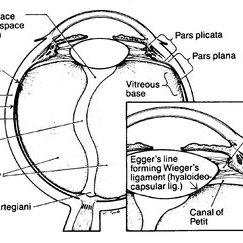 Classic Human Vitreous Anatomy
Sep 1 2020 by J. Sebag, MD, FACS, FRCOphth, FARVO
Schematic diagram of human vitreous anatomy depicting structures often named after anatomists. [Originally From Sang D: Embryology of the vitreous - congenital and developmental abnormalities. In: The Vitreous and Vitreoretinal Interface (CL Schepens, A Neetens, eds). Springer Verlag, New York, pg 20; reprinted in Sebag J: The Vitreous- Structure, Function, and Pathobiology, Springer-Verlag, New York, 1989; image © Springer Nature, reprinted with permission]
Condition/keywords: vitreous
-
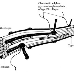 Human Vitreous Collagen Fibril
Sep 1 2020 by J. Sebag, MD, FACS, FRCOphth, FARVO
Schematic diagram of human vitreous collagen fibril showing the central core of hybrid types V/XI surrounded by type II collagen, the most prevalent type in both vitreous and joints. Type IX is located on the surface of the fibril where it can mediate interactions with other extracellular matrix components in the vitreous body. [From Bishop PN: The biochemical structure of mammalian vitreous. Eye 1996;10:664–70; reprinted in Sebag J: Vitreous – in Health & Disease. Springer, New York, 2014; image © Springer Nature, reprinted with permission]
Condition/keywords: collagen, vitreous, vitreous fibrils
-
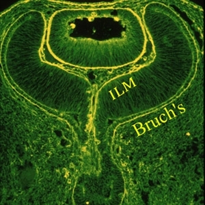 Human Embryonic Eye
Sep 1 2020 by J. Sebag, MD, FACS, FRCOphth, FARVO
Immunohistochemistry of a human fetal eye after invagination of the optic vesicle demonstrates basement membrane staining with fluorescein conjugated ABA lectin staining. Continuity of the membranes destined to be Bruch’s membrane and the Inner Limiting Membrane (ILM) of the retina is evident (see loops at upper aspects of this image), suggesting a common embryologic origin and similar composition. Thus, there may be important similarities during pathologies (especially neovascularization) at these two interfaces, which may provide insights into the process of pathologic cell migration and proliferation. Anterior to the ILM and behind the lens is the vascular primary vitreous. [Cover Photo - Sebag J and Hageman G: Interfaces. Farina Publishers, Rome, 2000; reprinted in Sebag J: Vitreous – in Health & Disease (J. Sebag, ed.) Springer, New York, 2014; image © Springer Nature, reprinted with permission]
Condition/keywords: embryonic eye
-
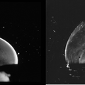 Vitreous Structure in Youth
Sep 1 2020 by J. Sebag, MD, FACS, FRCOphth, FARVO
Dark-field slit microscopy was performed on fresh, unfixed, post-mortem human eyes that had undergone dissection to peel off the sclera, choroid, and retina. The vitreous body remains attached to the anterior segment which is seen below, while the posterior pole is above in these images. These horizontal optical sections demonstrate intense light scattering by the posterior vitreous cortex and the remnant of the hyaloid artery destined to be Cloquet’s Canal (left image), but no other light scattering within the vitreous body in either the 33 GW human fetus (left image) or this 6 year-old child (right image). [from Sebag J: The Vitreous - Structure, Function, and Pathobiology. Springer-Verlag, New York, 1989, left image pg. 77; right image pg. 79; images © Springer Nature, reprinted with permission]
Condition/keywords: vitreous
-
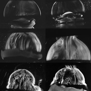 Age-Related Differences in the Structure of the Human Vitreous Body
Sep 1 2020 by J. Sebag, MD, FACS, FRCOphth, FARVO
Dark-field slit microscopy was performed on fresh, unfixed, post-mortem human eyes that had undergone dissection to peel off the sclera, choroid, and retina. The vitreous body remains attached to the anterior segment which is seen below, while the posterior pole is above in these images. The top panel demonstrates the absence of internal vitreous structures that scatter light in youth (left image from an 11 year-old girl, right image from a 14 year-old boy. The middle panel demonstrates light scattering from linear, fibrous structures that have an antero-posterior orientation with insertions into the vitreous base peripherally and the posterior vitreous cortex, typical in middle age (left image from a 56 year-old and right image from a 59 year-old). The bottom panel illustrates advance fibrous liquefaction in old age (88-year-old subject). [From Sebag J, Niemeyer M, Koss M: Anomalous PVD and vitreoschisis. In: Vitreous – in Health & Disease (J. Sebag, ed.) Springer, New York, 2014, pg. 245; image © Springer Nature, reprinted with permission]
Condition/keywords: vitreous
-
 Human Hyalocytes
Sep 1 2020 by J. Sebag, MD, FACS, FRCOphth, FARVO
LEFT: Dark-field slit microscopy was performed on this fresh, unfixed, post-mortem human eye that had undergone dissection to peel off the sclera, choroid, and retina. The posterior pole is imaged in this image with vitreous extruding out the prepapillary hole in the posterior vitreous cortex (small, to right) and the premacular dehiscence (larger, to left). Bright light scattering is seen from punctate structures with the posterior vitreous cortex, corresponding to hyalocytes distributed in a monolayer. RIGHT: Transmission electron microscopy of human hyalocyte in situ demonstrates embedding of the hyalocyte within the dense collagen matrix of the posterior vitreous cortex. Mi = microvilli; black C = collagen of posterior vitreous cortex; N = lobulated nucleus typical of mononuclear phagocytes; white C = dense marginal chromatin in nucleus; M = mitochondria; V = vacuoles; arrows = dense granule (original magnification = 11,670) [From Sebag J: The Vitreous - Structure, Function, and Pathobiology. Springer-Verlag, New York, 1989; right 48, left pg. 50 (images © Springer Nature, reprinted with permission); right image courtesy of J. L. Craft and D. M. Albert, MD, Harvard Medical School, Boston, MA]
Condition/keywords: hyalocytes
-
 Human Vitreous Base Structure
Sep 1 2020 by J. Sebag, MD, FACS, FRCOphth, FARVO
Dark-field slit microscopy was performed on fresh, unfixed, post-mortem human eyes that had undergone dissection to peel off the sclera, choroid, and retina. The vitreous body remains attached to the anterior segment which is seen below, while the posterior pole is above in these images. Left: specimen was tilted to reveal the posterior aspect of the lens (L) and the fibers of the vitreous base (arrow) splayed out to insert anterior and posterior to the ora serrata; Right: Anterior Loop of the vitreous base (see text). [From Sebag J: The Vitreous - Structure, Function, and Pathobiology. Springer-Verlag, New York, 1989, pp. 41 & 42; images © Springer Nature, reprinted with permission]
Condition/keywords: vitreous
-
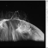
 Posterior Vitreous Detachment
Sep 1 2020 by J. Sebag, MD, FACS, FRCOphth, FARVO
Left: Preset lens biomicroscopy of PVD in the left eye of a subject with a widely dilated pupil. The detached posterior vitreous cortex is seen (arrows) as is the optic disc and retinal vasculature (upper left). (courtesy of C. L. Trempe MD, Harvard Medical School, Boston, MA) [Sebag J: Vitreous – in Health & Disease Springer, New York, 2014; image © Springer Nature, reprinted with permission] Right: B-scan ultrasonography of PVD images the detached posterior vitreous cortex with a visible Weiss Ring.
Condition/keywords: posterior vitreous detachment
-
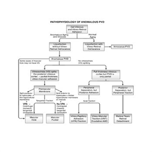 Pathophysiology of Anomalous PVD
Sep 1 2020 by J. Sebag, MD, FACS, FRCOphth, FARVO
This unifying concept of vitreo-retinopathies hypothesizes that the pathogenesis of several vitreoretinal diseases that were previously considered very disparate, are actually all manifestations of the same underlying pathophysiology – anomalous PVD. Note that vitreo-papillary adhesion (VPA) and traction can cause primary optic neuropathy, but might also play a role in facilitating/promoting cell migration and proliferation during pathologic neovascularization of the optic disc. Further, VPA seems to alter the vector of tangential forces exerted by a membrane, in some cases full-thickness posterior vitreous cortex and in some cases the outer layer of the posterior vitreous cortex left attached to the macula after vitreoschisis. While not all cases of macular holes have vitreoschisis, they feature vitreomacular adhesion and traction almost always with VPA. [From Sebag J: Anomalous PVD – a unifying concept in vitreo-retinal diseases. Graefe’s Arch Clin Exp Ophthalmol 2004;242:690-8 and Sebag J, Niemeyer M, Koss M: Anomalous PVD and vitreoschisis. In: Vitreous – in Health & Disease (J. Sebag, ed.) Springer, New York, 2014, pg. 252; image © Springer Nature, reprinted with permission]
Condition/keywords: pathology, peripheral vascular disease (PVD)
-
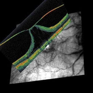 Vitreo-Macular Traction Syndrome
Sep 1 2020 by J. Sebag, MD, FACS, FRCOphth, FARVO
Combined OCT and Scanning laser ophthalmoscopy demonstrate separation of full-thickness posterior vitreous cortex (no vitreoschisis), but persistent adhesion centrally with significant detachment of the fovea. [from Sebag J: Vitreous – in Health & Disease (J. Sebag, ed.) Springer, New York, 2014; image © Springer Nature, reprinted with permission]
Condition/keywords: vitreomacular traction (VMT)
-
 Pathophysiology of Vitreoschisis
Sep 1 2020 by J. Sebag, MD, FACS, FRCOphth, FARVO
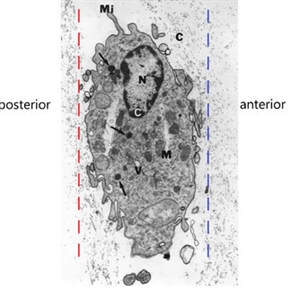
Transmission electron microscopy of human hyalocyte in situ demonstrates embedding within the dense collagen matrix of the posterior vitreous cortex. The retina was to the left (“posterior”) and the anterior segment was to the right (“anterior”). The red dashed line indicates the level of vitreoschisis split that might occur posterior to the level of the hyalocyte monolayer, leaving a thin, hypocellular membrane attached to the macula. The dashed blue line indicates the level of vitreoschisis split that might occur anterior to the level of the hyalocyte monolayer, leaving a thick, hypercellular membrane attached to the macula. The former is more likely to present as macular hole, while the latter as macular pucker (see Figure 12). Mi = microvilli; black C = collagen of posterior vitreous cortex; N = lobulated nucleus typical of mononuclear phagocytes; white C = dense marginal chromatin in nucleus; M = mitochondria; V = vacuoles; arrows = dense granule (original magnification = 11,670) [Modified from Sebag J: Anomalous PVD – a unifying concept in vitreo-retinal diseases. Graefe’s Arch Clin Exp Ophthalmol 2004;242:690-8 and Sebag J, Niemeyer M, Koss M: Anomalous PVD and vitreoschisis. In: Vitreous – in Health & Disease (J. Sebag, ed.) Springer, New York, 2014, pg. 252]
Condition/keywords: pathology, vitreoschisis
-
 Senescent Human Vitreous Structure
Sep 1 2020 by J. Sebag, MD, FACS, FRCOphth, FARVO
Dark-field slit microscopy was performed on a fresh, unfixed, post-mortem human eye from an 88 year-old subject. The eye had dissection to peel off the sclera, choroid, and retina. The vitreous body remains attached to the anterior segment which is seen below, while the posterior pole is above in this image. Significant degenerative fibrous liquefaction is evident. Lacunae can be seen (arrows) that do not scatter light because they are primarily filled with liquid vitreous that is mostly hyaluronan and water, with little collagen to scatter light. Drug injection into a lacuna would likely have very different distribution than injection into an area of gel vitreous. [From Sebag J: The Vitreous - Structure, Function, and Pathobiology. Springer-Verlag, New York, 1989 (image © Springer Nature, reprinted with permission); reprinted in Sebag J: Vitreous in AMD therapy – the medium is the message (Guest Editorial). Retina 2015;35(9):1715-18]
Condition/keywords: vitreous
A project from the American Society of Retina Specialists