Search results (12 results)
-
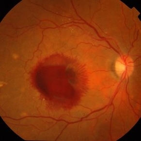
 Ruptured Retinal Artery Macroaneurysm
Jun 18 2024 by KANWALJEET HARJOT MADAN, M.S. (Ophthalmology); FAICO (Vitreous - Retina)
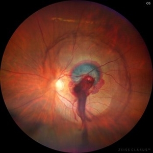
This is a fundus photo depicting ruptured Retinal Artery Macroaneurysm (RAM) in the left eye of a 63 years old female. RAM is an acquired saccular or fusiform dilatation of the retinal arterioles that usually occur within the first three orders of bifurcation. The Superotemporal artery is the most common location. RAM may be asymptomatic or cause a number of complications such as macular edema, serous macular detachment, and hemorrhages.
Photographer: Dr Kanwaljeet Harjot Madan
Condition/keywords: Haemorrhage, macroaneurysm, retinal arteriole
-
 Perforating Ocular Trauma and Choroidal Rupture due to Shotgun Pellet
Mar 31 2022 by Franco Benvenuto, MD
Fundus photograph of a 17-year-old with shotgun injuries with numerous metal pellets in the chest, neck, brain, and face. Fundus exploration showed the left globe with posterior-inferior eye rupture, vitreous hemorrhages and choroidal rupture.
Photographer: Franco Benvenuto, Universidad de Buenos Aires, Argentina. Universidad de Guadalajara, México.
Condition/keywords: choroidal rupture, penetrating trauma, shotgun
-
 Blunt Ocular Trauma Due to Firework Injury
Jun 9 2020 by Brittany Rota
Ultra- widefield pseudocolor image of an 18-year-old male with blunt ocular trauma in the right eye due to a firework injury. The patient presented with commotio retinae (sclopteria), an acute vitreous hemorrhage, choroidal rupture, and a subretinal hemorrhage. The referring physician performed surgery on the lateral rectus muscle which was macerated but not severed, and several orbital fibrous foreign bodies were removed from the posterior orbit. The globe was intact. There is no evidence of retinal tear in the region of sclopetaria; however, there is complete necrosis of the temporal peripheral choroid and retina. The vitreous hemorrhage was slowly clearing on his exam 6-9-2020. The patient is developing subretinal fibrosis. The physician is concerned about the choroidal rupture that is visible through the submacular hemorrhage. There is one rupture that appears to course directly under the fovea. The physician states that if this is the case, his vision most likely will be 20/200 or worse. His vision was hand motion in all fields except nasally, which he was unable to see hand motion at his visit on 6-9-2020.
Photographer: Brittany Rota
Imaging device: Optos California
Condition/keywords: blunt trauma, choroidal rupture, commotio retinae, fibrosis, firework injury, fundus photograph, hand motion, necrotizing retina, Optos, pseudocolor, subretinal hemorrhage, vitreous hemorrhage
-
 Surfboard to OS - Choroidal Rupture, Retinal and Choroidal Avulsion, Retinal Detachment
May 20 2020 by Theodore Leng, MD, MS, FASRS
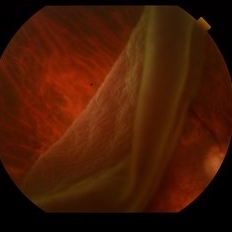
33-year-old male who sustained a surfboard to the left orbit with orbital fractures, choroidal rupture, nasally and retinal and choroidal avulsion nasally. He also had a horseshoe retinal tear superotemorally and a resultant rhegmatogenous retinal detachment.
Condition/keywords: choroidal avulsion, choroidal rupture, retinal avulsion, retinal detachment with retinal defect
-
 Ruptured Macroaneurysm
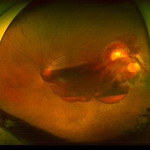
May 22 2019 by Nichole Lewis
FA of a 91-year-old woman with a ruptured macroaneurysm, intraretinal hemorrhage and subretinal hemorrhage. VA 20/400.
Photographer: Nichole Lewis
Condition/keywords: intraretinal hemorrhage, ruptured macroaneurysm, subretinal hemorrhage
-
 Penetrating Trauma with Retinal Detachment
Apr 30 2019 by Olivia Rainey
Ultra-wide field pseudocolor image of a 39-year-old female with penetrating trauma resulting in a retinal detachment with an intraretinal hemorrhage affecting the left eye. Patient was struck with a champagne glass in October of 2018, which lacerated the eyelid and globe. Patient was "seeing red" when she first came to the office and after multiple surgeries she was seeing 20/20 at her last check in April 2019.
Photographer: Olivia Rainey
Imaging device: Optos
Condition/keywords: hemorrhage, left eye, Optos, penetrating trauma, ruptured globe, ultra-wide field imaging
-
 Retinal Detachment
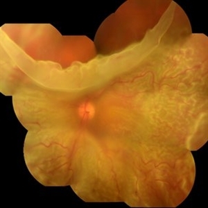
Oct 25 2018 by Graciela Nahuelquín Ríos
A 45-year-old patient reported a blow to the right eye 1 month ago, and a week ago he presented with low visual acuity. In retinal mapping and background color photography retinal detachment with giant rupture in temporal arch.
Photographer: Lic. TM. Graciela Nahuelquín Ríos
Imaging device: TRC-50DX - Topcon
-
 Multiple Retinal Lesions Secondary to Blunt Trauma
Jun 19 2018 by Somnath Chakraborty, MD
A montage of the right eye of a 15-year-old boy, who was struck by a football. The image shows multiple choroidal ruptures in the macular area, with sub-retinal blood and multiple, large retinal tears temporally. There is also an area of juxtapapillary, pigmentary changes.
Photographer: Saptarshi Mehta, Retina Institute of Bengal
Condition/keywords: blunt trauma, choroidal rupture, giant retinal tear, subretinal hemorrhage
-
 Retinal Detachment
Feb 8 2018 by JEFFERSON R SOUSA, Tecg.º (Biomedical Systems Technology)
The male patient attended the clinic with low vision. In the retinal and retinal mapping examination, important fudoscopical alterations were observed. Full retinal detachment with Giant rupture in upper temporal arch.
Photographer: JEFFERSON R SOUSA - Study Center and Ophthalmological Research Dr. Andre M V Gomes, Institute Dr. Suel Abujamra São Paulo-Brazil
Imaging device: Fundus camera Topcon TRC-50 DX, Imaginet 5.0, campo de 50 graus. Flash 36 / Mosaic with 16 images.
Condition/keywords: retinal in rupture
-
 Traumatic Choroidal Rupture
Jun 11 2017 by Bastián Schmidt Arias
Fundus photograph of an 28-year-old men with a traumatic choroidal rupture. Visual acuity is CF 30 cm.
Photographer: Bastian Schmidt
Imaging device: TRC-50DX - Topcon
Condition/keywords: choroidal rupture, fundus photograph
-
 Macroaneurysm
Apr 1 2017 by Manish Nagpal, MD, FRCS (UK), FASRS
Case of a ruptured macroaneurysm with subhyaloid and subretinal blood.
Photographer: Avijit Vishnoi
Condition/keywords: macroaneurysm, ruptured macroaneurysm
-
 RAMA
RAMA
Jun 20 2016 by John S. King, MD
RAMA with 2 w co decreased vision; htn, afib using anticoag; light laser applied; 20/400.
Condition/keywords: ruptured macroaneurysm
 Loading…