Search results (274 results)
-
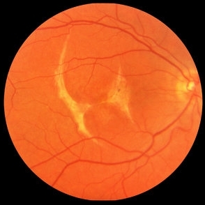
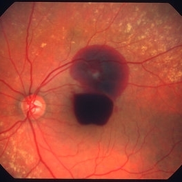 Ruptured retinal arterial macroaneurysm
Jan 11 2013 by Alex P. Hunyor, MD
Retinal arterial macroaneurysm with subretinal and preretinal hemorrhage
Condition/keywords: retinal arterial macroaneurysm
-
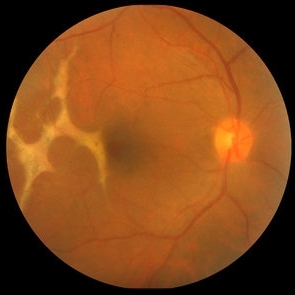
 Choroidal Rupture with Subretinal Hemorrhage
Oct 1 2012 by Jeffrey G. Gross, MD, FASRS
Choroidal rupture with subretinal hemorrhage.
Condition/keywords: choroidal rupture, subretinal hemorrhage
-
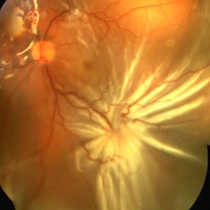
 Choroidal Rupture with Extensive Subretinal Hemorrhage HM
Oct 1 2012 by Jeffrey G. Gross, MD, FASRS
Choroidal rupture with extensive subretinal hemorrhage HM.
Condition/keywords: choroidal rupture, HM, subretinal hemorrhage
-
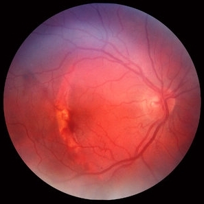
 Choroidal Rupture Across Macula
Oct 23 2012 by Larry Halperin, MD
Choroidal rupture across macula
Condition/keywords: choroidal rupture, macula
-
 Juxtafoveal Choroidal Neovascularization Secondary to Choroidal Rupture
Aug 30 2012 by Young Hee Yoon, MD, PhD
SD-OCT image of a 14-year-old boy with a history of blunt trauma to his left eye 9 months ago. Best-corrected visual acuity remained at 20/30.
Photographer: Soo Hyun Cho, Asan Medical Center
Imaging device: HHeidelberg Spectralis OCTI/ version 1.7.0.0
Condition/keywords: choroidal rupture, juxtafoveal choroidal neovascularization (CNV)
-
 Juxtafoveal Choroidal Neovascularization Secondary to Choroidal Rupture
Aug 30 2012 by Young Hee Yoon, MD, PhD
Fundus photograph of a 14-year-old boy with a history of blunt trauma to his left eye 9 months ago. Best-corrected visual acuity remained at 20/30.
Photographer: Heon Eui Hong, Asan Medical Center
Imaging device: Canon CR-DGI / Version 5.1.2
Condition/keywords: choroidal rupture, juxtafoveal choroidal neovascularization (CNV)
-
 Acute Posterior Vitreous Detachment
Nov 9 2012 by Norman Byer
This large and complicated retinal tear in a 51-year-old man resulted from an acute posterior vitreous detachment which concentrated its tractional forces around this area of lattice degeneration. Because of the powerful traction, there is an additional central tear splitting the large retinal flap and almost severing one of its arms. The traction was strong enough to completely rupture the blood vessel just to the left of the flap. Marking the ruptured peripheral end of the blood vessel is a yellow depigmented thrombus.
Condition/keywords: acute posterior vitreous detachment, depigmented thrombus, lattice degeneration, retinal tear, tractional retinal detachment
-
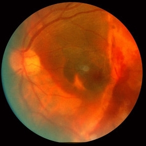
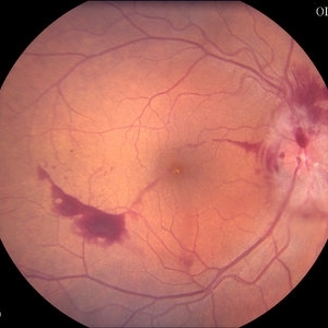
 Traumatic Choroidal Rupture
Jun 11 2017 by Bastián Schmidt Arias
Fundus photograph of an 28-year-old men with a traumatic choroidal rupture. Visual acuity is CF 30 cm.
Photographer: Bastian Schmidt
Imaging device: TRC-50DX - Topcon
Condition/keywords: choroidal rupture, fundus photograph
-
 Bruch’s membrane rupture
Jan 11 2013 by Hyung-Woo Kwak, MD
An area of Bruch’s membrane rupture involving the fovea is seen on color photograph (left).
Photographer: Misook Lee, Kyung Hee Univsersity Hospital, Seoul
Imaging device: Zeiss f 450 plus
Condition/keywords: myopic choroidal neovascularization (CNV)
-
 Choroidal Hemangioma
Oct 20 2012 by Hyung-Woo Kwak, MD
Fundus, ICG, and OCT examination showed a typical chorioretinal scar lying concentric to the optic disc. Typical choroidal rupture was performed after intravitreal gas injection under the diagnosis of submacular hemorrhage caused by trauma, after the absorption of submacular hemorrhage
Condition/keywords: chorioretinal scar, choroidal rupture, submacular hemorrhage
-
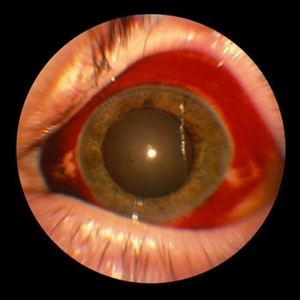 Subconjunctival Hemorrhage
Sep 20 2012 by Jeffrey G. Gross, MD, FASRS
Subconjunctival hemorrhage, trauma in eye with choroidal rupture HM
Condition/keywords: choroidal rupture, subconjunctival hemorrhage
-
 Retinitis Sclopetaria, 6 months later
Jun 29 2018 by Gareth Lema, MD, PhD
Retinitis sclopetaria has resolved. There are multiple, large choroidal ruptures and subretinal scarring.
Photographer: Flaum Eye Institute, University of Rochester, Rochester, NY
Condition/keywords: blunt trauma, chorioretinitis sclopetaria
-
 Choroidal Rupture
-
 Choroidal Rupture
Oct 16 2012 by Jeffrey G. Gross, MD, FASRS
Choroidal rupture, s/p PPV, with TPA, and gas pneumatic displacement.
Condition/keywords: 10L technique, choroidal rupture, gas pneumatic displacement
-
 Starfold in Proliferative Vitreoretinopathy
Sep 11 2014 by Thomas A. Ciulla, MD, MBA, FASRS
This patient underwent repair of a rupture globe, extraction of an intraocular foreign body, vitrectomy, laser, and silicone oil placement. When he returned for follow up, he was noted to have a recurrent shallow retinal detachment under the silicone oil, with a prominent star fold at the inferior temporal arcade.
Photographer: Thomas Steele
Condition/keywords: proliferative vitreoretinopathy (PVR)
-
 Choroidal Rupture through Fovea
Oct 1 2012 by Jeffrey G. Gross, MD, FASRS
Choroidal rupture through fovea.
Condition/keywords: choroidal rupture, fovea
-
 Bruch’s membrane rupture
Jan 11 2013 by Hyung-Woo Kwak, MD
An area of Bruch’s membrane rupture involving the fovea is seen on indocyanine green angiography: late phase (right).
Photographer: Misook Lee, Kyung Hee Univsersity Hospital, Seoul
Imaging device: Zeiss f 450 plus
Condition/keywords: Bruch's membrane, myopic choroidal neovascularization (CNV)
-
 Juxtafoveal Choroidal Neovascularization Secondary to Choroidal Rupture
Aug 30 2012 by Young Hee Yoon, MD, PhD
Fluorescence Angiography (FA) image of a 14-year-old boy with a history of blunt trauma to his left eye 9 months ago. Best-corrected visual acuity remained at 20/30.
Photographer: Heon Eui Hong, Asan Medical Center
Imaging device: HHeidelberg HRA II/ version 1.7.0.0
Condition/keywords: choroidal rupture, juxtafoveal choroidal neovascularization (CNV)
-
 Tractional Retinal Tear
Nov 9 2012 by Norman Byer
This is the same lesion and shows the free operculum in better focus. This is an unusual location for a tractional retinal tear, and the increased mobility of the detached vitreous in the posterior part of the eye may have been a factor leading to the complete rupture of this retinal flap.
Condition/keywords: detached vitreous, free operculum, tractional retinal tear
-
 Ruptured Globe Trauma
Jul 11 2013 by Jason S. Calhoun
Young male who got hit with a baseball ended up with orbital floor fracture and ruptured globe.
Photographer: Jason S. Calhoun, Department of Ophthalmology, Mayo Clinic Jacksonville, Florida
Condition/keywords: blunt trauma
-
 Macroaneurysm - OCT
Oct 5 2013 by Roy Schwartz, MD
Right eye of a 84-year-old female with a macroaneurysm.
Photographer: Galit Yair-Pur
Condition/keywords: macroaneurysm, optical coherence tomography (OCT), ruptured macroaneurysm
-
 Juxtafoveal Choroidal Neovascularization Secondary to Choroidal Rupture
Aug 30 2012 by Young Hee Yoon, MD, PhD
Indocyanine Green Angiography (ICGA) image of a 14-year-old boy with a history of blunt trauma to his left eye 9 months ago. Best-corrected visual acuity remained at 20/30.
Photographer: Heon Eui Hong, Asan Medical Center
Imaging device: HHeidelberg HRA II/ version 1.7.0.0
Condition/keywords: choroidal rupture, juxtafoveal choroidal neovascularization (CNV)
-
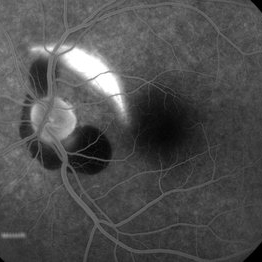 Choroidal rupture and peripapillary hemorrhage - FA
Jan 26 2013 by Roy Schwartz, MD
A 36-year-old male presented to the ER after blunt trauma to his left eye. On FA a chroidal rupture (hyperfluorescent area) was seen as well as peripapillary hemorrhage (hypofluorescent).
Photographer: Galit Yair-Pur
Condition/keywords: choroidal rupture, peripapillary hemorrhage
-
 Macroaneurysm - Fundus Image
Oct 5 2013 by Roy Schwartz, MD
Right eye of a 84-year old female with a macroaneurysm.
Photographer: Galit Yair-Pur
Condition/keywords: macroaneurysm, ruptured macroaneurysm
-
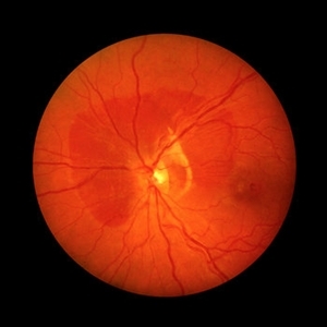
 Diabetic Papillitis
Jun 28 2013 by Jason S. Calhoun
Patient was about to undergo surgery for CNS aneurysm. Patient woke up with little spots which never cleared up. Patients VA was 20/30-OD and 20/40-OS. Both eyes appeared to have disc edema with hemorrhages in the right eye. Ordered a CT of the brain to make sure the aneurysm didn't ruptured.
Photographer: Jason S. Calhoun, Mayo Clinic Jacksonville, Florida
Imaging device: TOPCON TRC 50-EX
Condition/keywords: diabetic mellitus
 Loading…